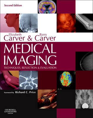
Medical Imaging: Techniques, Reflection & Evaluation
Churchill Livingstone (Verlag)
978-0-7020-3933-1 (ISBN)
- Titel erscheint in neuer Auflage
- Artikel merken
Medical Imaging has been revised and updated to reflect the current role and responsibilities of the radiographer, a role that continues to extend as the 21st century progresses. This comprehensive book covers the full range of medical imaging methods/techniques which all students and professionals must understand, and discusses them related to imaging principles, radiation dose, patient condition, body area and pathologies.
There is comprehensive, up-to-date, referencing for all chapters, with full image evaluation criteria and a systematic approach to fault recognition for all radiographic projections. Highly respected editors, Elizabeth and Barry Carver, have brought together an impressive team of contributing authors, comprising academic, radiographer and radiologist clinical experts. NEW TO THIS EDITION
Full colour, including approximately 200 new colour photographs.All techniques have been updated to reflect the use of digital image receptors. All chapters have been updated to reflect current practice, eg CT colonoscopy is now included as part of GI imaging; the nuclear medicine chapter now introduces hybrid imaging; the genitourinary chapter now reflects the use of ultrasound and CT.'The authors have been comprehensive, thorough and innovative. This well-presented book should be adopted by Schools of Diagnostic Imaging in Europe and elsewhere and be a constant companion to the reflective radiographic practitioner.'
From the foreword to the first edition by Patrick Brennan.
Medical Imaging has been revised and updated to reflect the current role and responsibilities of the radiographer, a role that continues to extend as the 21st century progresses. This comprehensive book covers the full range of medical imaging methods/techniques which all students and professionals must understand, and discusses them related to imaging principles, radiation dose, patient condition, body area and pathologies.
There is comprehensive, up-to-date, referencing for all chapters, with full image evaluation criteria and a systematic approach to fault recognition for all radiographic projections. Highly respected editors, Elizabeth and Barry Carver, have brought together an impressive team of contributing authors, comprising academic, radiographer and radiologist clinical experts.
Full colour, including approximately 200 new colour photographs.All techniques have been updated to reflect the use of digital image receptors. All chapters have been updated to reflect current practice, eg CT colonoscopy is now included as part of GI imaging; the nuclear medicine chapter now introduces hybrid imaging; the genitourinary chapter now reflects the use of ultrasound and CT.
Section 1 Imaging principles
1. Digital imaging
2. Film-screen imaging
3. Exposure factors, manipulation and dose
Section 2 Skeletal radiography
4. Introduction to skeletal, chest and abdominal radiography
5. Fingers, hand and wrist
6. Forearm, elbow and humerus
7. The shoulder girdle
8. Foot, toes, ankle, tibia and fibula
9. Knee and femur
10. Pelvis and hips
11. Cervical spine
12. Thoracic spine
13. Lumbar spine
14. Sacrum and coccyx
15. Thoracic skeleton
16. Principles of radiography of the head
17. Cranial vault
18. Facial bones
19. Paranasal sinuses
20. Specialised projections of the skull
21. Dental radiography
22. Orthopantomography and cephalometry
Section 3 Chest and abdomen
23. Chest and thoracic contents
24. Abdomen
Section 4 Accident and emergency
25. Accident and emergency
Section 5 Breast imaging
26. Breast imaging
Section 6 Paediatric imaging
27. Paediatric imaging in general radiography
Section 7 Contrast studies
28. Contrast media
29. Gastrointestinal tract
30. Accessory organs of the gastrointestinal tract
31. Investigations of the genitourinary tract
32. Cardiovascular system
33. Vascular imaging of the head and neck
34. Intervention and therapeutic procedures
Section 8 Additional imaging methods
35. Computed tomography
36. Magnetic Resonance Imaging
37. Nuclear Medicine Imaging
38. Ultrasound
Glossary of radiographic terms
Index
| Erscheint lt. Verlag | 16.7.2012 |
|---|---|
| Verlagsort | London |
| Sprache | englisch |
| Maße | 276 x 219 mm |
| Gewicht | 1860 g |
| Themenwelt | Studium ► 2. Studienabschnitt (Klinik) ► Anamnese / Körperliche Untersuchung |
| ISBN-10 | 0-7020-3933-0 / 0702039330 |
| ISBN-13 | 978-0-7020-3933-1 / 9780702039331 |
| Zustand | Neuware |
| Haben Sie eine Frage zum Produkt? |
aus dem Bereich


