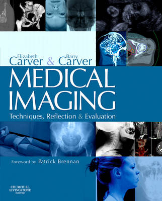
Medical Imaging
Churchill Livingstone (Verlag)
978-0-443-06212-4 (ISBN)
- Titel erscheint in neuer Auflage
- Artikel merken
This book provides in one concise volume the entire range of radiographic positioning and techniques - the fundamental concepts of radiography which all students and professionals must understand. Uniquely, it combines this essential core knowledge with a reflective approach. The editors have brought together contributions from radiographers, radiography lecturers, radiologists and other experts from the commercial sector of medical imaging, all selected for their clinical and academic expertise. The authors have been comprehensive, thorough and innovative. This well-presented book should be adopted by Schools of Diagnostic Imaging in Europe and elsewhere and be a constant companion to the reflective radiographic practitioner. (From the foreword by Patrick Brennan)
Section 1 Imaging principles 1.Film/screen imaging Susan Penelope Nash 2.Digital imaging Philip Cosson 3.Density, contrast and unsharpness, exposure factors and dose Barry Carver Section 2 Skeletal radiography 4.Introduction to Sections 2 and 3: Skeletal chest and abdominal radiography Elizabeth Carver and Barry Carver 5.Fingers, hand and wrist Elizabeth Carver 6.Forearm, elbow and humerus Elizabeth Carver 7.The shoulder girdle Linda Williams 8.Foot, toes, ankle, tibia and fibula Linda Williams 9.Knee and femur Linda Williams 10.Pelvis and hips Linda Williams 11.Cervical spine Barry Carver 12.Thoracic spine Linda Williams 13.Lumbar spine Margo McBride 14.Sacrum and coccyx Elizabeth Carver 15.Thoracic skeleton Elizabeth Carver 16.Principles of radiography of the head Elizabeth Carver 17.Basic isocentric radiography Amanda J. Royle 18.Cranial vault Barry Carver and Amanda.J. Royle 19.Face (facial bones) Elizabeth Carver and Amanda J. Royle 20.Paranasal sinuses Elizabeth Carver and Amanda J. Royle 21.Specialised skull Elizabeth Carver and Amanda J. Royle 22.Intra-oral dental radiography Elizabeth Carver 23.Orthopantomography Elizabeth Carver 24. Cephalometry Elizabeth Carver Section 3 Chest and abdomen 25.Chest and thoracic contents Elizabeth Carver 26.Abdomen Elizabeth Carver Section 4 A & E imaging 27.Accident and emergency Jonathan McConnell and Elizabeth Carver Section 5 Mammography 28.Mammography: introduction and rationale Julie Burnage 29.Mammography technique Julie Burnage 30.Mammography ultrasound Julie Burnage 31.Mammography localisation Julie Burnage Section 6 Paediatric imaging 32.Paediatric imaging Tim Palarm and Michael Scriven Section 7 Contrast studies 33.Contrast agents Susan Cutler 34.Investigations of GI tract Joanne Rudd and Darren Wood 35.Accessory organs of GI tract Darren Wood 36.Investigations of genito-urinary tract Catherine Williams and Elizabeth Carver 37.Cardiovascular system Mark Cowling 38.Interventional and vascular procedures Mark Cowling 39.Head and neck Patricia Fowler and Andrew Layt Section 8 Comparative imaging 40.CT Barry Carver 41.MRI John Talbot 42.Radionuclide imaging David Jones and Julian Macdonald 43.Ultrasound Mike Stocksley and Rita Phillips Index
| Erscheint lt. Verlag | 18.5.2006 |
|---|---|
| Zusatzinfo | Approx. 800 illustrations |
| Verlagsort | London |
| Sprache | englisch |
| Maße | 219 x 276 mm |
| Gewicht | 1000 g |
| Themenwelt | Medizinische Fachgebiete ► Radiologie / Bildgebende Verfahren ► Radiologie |
| ISBN-10 | 0-443-06212-9 / 0443062129 |
| ISBN-13 | 978-0-443-06212-4 / 9780443062124 |
| Zustand | Neuware |
| Haben Sie eine Frage zum Produkt? |
aus dem Bereich


