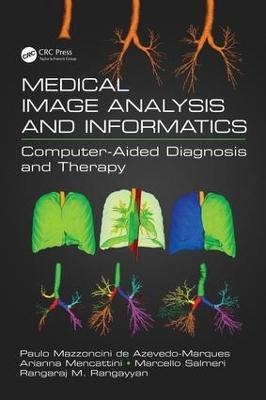
Medical Image Analysis and Informatics
Crc Press Inc (Verlag)
978-1-4987-5319-7 (ISBN)
With the development of rapidly increasing medical imaging modalities and their applications, the need for computers and computing in image generation, processing, visualization, archival, transmission, modeling, and analysis has grown substantially. Computers are being integrated into almost every medical imaging system. Medical Image Analysis and Informatics demonstrates how quantitative analysis becomes possible by the application of computational procedures to medical images. Furthermore, it shows how quantitative and objective analysis facilitated by medical image informatics, CBIR, and CAD could lead to improved diagnosis by physicians. Whereas CAD has become a part of the clinical workflow in the detection of breast cancer with mammograms, it is not yet established in other applications. CBIR is an alternative and complementary approach for image retrieval based on measures derived from images, which could also facilitate CAD. This book shows how digital image processing techniques can assist in quantitative analysis of medical images, how pattern recognition and classification techniques can facilitate CAD, and how CAD systems can assist in achieving efficient diagnosis, in designing optimal treatment protocols, in analyzing the effects of or response to treatment, and in clinical management of various conditions. The book affirms that medical imaging, medical image analysis, medical image informatics, CBIR, and CAD are proven as well as essential techniques for health care.
Paulo Mazzoncini de Azevedo Marques is Associate Professor of Biomedical Informatics and Medical Physics. He is a Fellow of IEEE: Arianna Menattini, is in the Dept. of Electronic Engineering, at the Univ. of Rome Tor Vergata and is an active researcher in medical image analysis; Marcello Salmeri is an Associate Professor at the Univ. of Rome Tor Vergata; Rangaraj M. Rangayyan is an IEEE Fellow, Fellow AIMBE and SPIE. He ia extremely active in Computer Aided Diagnostic analysis and therapies for a number of different maladies.
Foreword on CAD: Its Past, Present, and Future
Preface
Acknowledgment
Contributors
1 Segmentation and Characterization of White Matter Lesions in FLAIR Magnetic Resonance Imaging
2 Computer-Aided Diagnosis with Retinal Fundus Images
3 Computer-Aided Diagnosis of Retinopathy of Prematurity in Retinal Fundus Images
4 Automated OCT Segmentation for Images with DME
5 Computer-Aided Diagnosis with Dental Images
6 CAD Tool and Telemedicine for Burns
7 CAD of Cardiovascular Diseases
8 Realistic Lesion Insertion for Medical Data Augmentation
9 Diffuse Lung Diseases (Emphysema, Airway and Interstitial Lung Diseases)
10 Computerized Detection of Bilateral Asymmetry
11 Computer-Aided Diagnosis of Breast Cancer with Tomosynthesis Imaging
12 Computer-Aided Diagnosis of Spinal Abnormalities
13 CAD of GI Diseases with Capsule Endoscopy
14 Texture-Based Computer-Aided Classification of Focal Liver Diseases using Ultrasound Images
15 CAD of Dermatological Ulcers (Computational Aspects of CAD for Image Analysis of Foot and Leg Dermatological Lesions)
16 In Vivo Bone Imaging with Micro-Computed Tomography
17 Augmented Statistical Shape Modeling for Orthopedic Surgery and
Rehabilitation
18 Disease-Inspired Feature Design for Computer-Aided Diagnosis of Breast Cancer Digital Pathology Images
19 Medical Microwave Imaging and Analysis
20 Making Content-Based Medical Image Retrieval Systems for Computer-Aided Diagnosis: From Theory to Application
21 Health Informatics for Research Applications of CAD
Concluding Remarks
Index
| Erscheinungsdatum | 24.11.2017 |
|---|---|
| Zusatzinfo | 119 Illustrations, color; 101 Illustrations, black and white |
| Verlagsort | Bosa Roca |
| Sprache | englisch |
| Maße | 178 x 254 mm |
| Gewicht | 1383 g |
| Themenwelt | Medizin / Pharmazie ► Medizinische Fachgebiete ► Radiologie / Bildgebende Verfahren |
| Medizin / Pharmazie ► Physiotherapie / Ergotherapie ► Orthopädie | |
| Studium ► 2. Studienabschnitt (Klinik) ► Anamnese / Körperliche Untersuchung | |
| Naturwissenschaften ► Physik / Astronomie ► Angewandte Physik | |
| Technik ► Medizintechnik | |
| Technik ► Umwelttechnik / Biotechnologie | |
| ISBN-10 | 1-4987-5319-1 / 1498753191 |
| ISBN-13 | 978-1-4987-5319-7 / 9781498753197 |
| Zustand | Neuware |
| Informationen gemäß Produktsicherheitsverordnung (GPSR) | |
| Haben Sie eine Frage zum Produkt? |
aus dem Bereich


