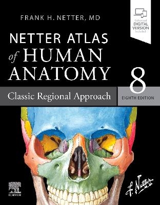
Netter Atlas of Human Anatomy: Classic Regional Approach
Elsevier - Health Sciences Division (Verlag)
978-0-323-68042-4 (ISBN)
- Presents world-renowned, superbly clear views of the human body from a clinical perspective, with paintings by Dr. Frank Netter as well as Dr. Carlos A. G. Machado, one of today's foremost medical illustrators.
- Content guided by expert anatomists and educators: R. Shane Tubbs, Paul E. Neumann, Jennifer K. Brueckner-Collins, Martha Johnson Gdowski, Virginia T. Lyons, Peter J. Ward, Todd M. Hoagland, Brion Benninger, and an international Advisory Board.
- Offers region-by-region coverage, including muscle table appendices at the end of each section and quick reference notes on structures with high clinical significance in common clinical scenarios.
- Contains new illustrations by Dr. Machado including clinically important areas such as the pelvic cavity, temporal and infratemporal fossae, nasal turbinates, and more.
- Features new nerve tables devoted to the cranial nerves and the nerves of the cervical, brachial, and lumbosacral plexuses.
- Uses updated terminology based on the second edition of the international anatomic standard, Terminologia Anatomica, and includes common clinically used eponyms.
- Enhanced eBook version included with purchase. Your enhanced eBook allows you to access all of the text, figures, and references from the book on a variety of devices.
- Provides access to extensive digital content: every plate in the Atlas?and over 100 bonus plates including illustrations from previous editions?is enhanced with an interactive label quiz option and supplemented with "Plate Pearls" that provide quick key points of the major themes of each plate. Digital content also includes over 300 multiple choice questions and other learning tools.
Also available, alternative versions of the 8th Edition:
Netter Atlas of Human Anatomy: A Systems Approach-Same content as the classic regional approach, but organized by organ systems.
Netter Atlas of Human Anatomy: Classic Regional Approach-hardback Professional Edition with downloadable image bank for personal use.
Netter Atlas of Human Anatomy: Classic Regional Approach with Latin terminology
Frank H. Netter was born in New York City in 1906. He studied art at the Art Students League and the National Academy of Design before entering medical school at New York University, where he received his Doctor of Medicine degree in 1931. During his student years, Dr. Netter's notebook sketches attracted the attention of the medical faculty and other physicians, allowing him to augment his income by illustrating articles and textbooks. He continued illustrating as a sideline after establishing a surgical practice in 1933, but he ultimately opted to give up his practice in favor of a full-time commitment to art. After service in the United States Army during World War II, Dr. Netter began his long collaboration with the CIBA Pharmaceutical Company (now Novartis Pharmaceuticals). This 45-year partnership resulted in the production of the extraordinary collection of medical art so familiar to physicians and other medical professionals worldwide. Icon Learning Systems acquired the Netter Collection in July 2000 and continued to update Dr. Netter's original paintings and to add newly commissioned paintings by artists trained in the style of Dr. Netter. In 2005, Elsevier Inc. purchased the Netter Collection and all publications from Icon Learning Systems. There are now over 50 publications featuring the art of Dr. Netter available through Elsevier Inc.
Dr. Netter's works are among the finest examples of the use of illustration in the teaching of medical concepts. The 13-book Netter Collection of Medical Illustrations, which includes the greater part of the more than 20,000 paintings created by Dr. Netter, became and remains one of the most famous medical works ever published. The Netter Atlas of Human Anatomy, first published in 1989, presents the anatomic paintings from the Netter Collection. Now translated into 16 languages, it is the anatomy atlas of choice among medical and health professions students the world over.
The Netter illustrations are appreciated not only for their aesthetic qualities, but, more importantly, for their intellectual content. As Dr. Netter wrote in 1949 "clarification of a subject is the aim and goal of illustration. No matter how beautifully painted, how delicately and subtly rendered a subject may be, it is of little value as a medical illustration if it does not serve to make clear some medical point." Dr. Netter's planning, conception, point of view, and approach are what inform his paintings and what make them so intellectually valuable.
Frank H. Netter, MD, physician and artist, died in 1991.
Section 1: Introduction
Section 2: Head and Neck
Section 3: Back and Spinal Cord
Section 4: Thorax
Section 5: Abdomen
Section 6: Pelvis and Perineum
Section 7: Upper Limb
Section 8: Lower Limb
| Erscheinungsdatum | 12.04.2022 |
|---|---|
| Reihe/Serie | Netter Basic Science |
| Zusatzinfo | Approx. 556 illustrations (556 in full color); Illustrations, unspecified |
| Verlagsort | Philadelphia |
| Sprache | englisch |
| Maße | 224 x 284 mm |
| Gewicht | 2420 g |
| Einbandart | kartoniert |
| Themenwelt | Studium ► 1. Studienabschnitt (Vorklinik) ► Anatomie / Neuroanatomie |
| ISBN-10 | 0-323-68042-9 / 0323680429 |
| ISBN-13 | 978-0-323-68042-4 / 9780323680424 |
| Zustand | Neuware |
| Informationen gemäß Produktsicherheitsverordnung (GPSR) | |
| Haben Sie eine Frage zum Produkt? |
aus dem Bereich


