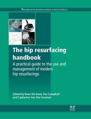
The Hip Resurfacing Handbook
Woodhead Publishing Ltd (Verlag)
978-1-84569-948-2 (ISBN)
Hip resurfacing arthroplasty (HRA) using metal-on-metal bearings is an established but specialised technique in joint surgery. Based on the experience of leading experts in the field, The hip resurfacing handbook provides a comprehensive reference for all aspects of this important procedure.The first part of the book reviews and compares all the major hip resurfacing prostheses, their key design features, relevant surgical techniques and clinical results. Part two discusses clinical follow-up of the hip resurfacing patient, including pre- and post-operative examination, acoustic phenomena and rehabilitation. It also covers the use of techniques such as radiography and metal ion measurement, as well as bone scans, ultrasound, CT, MRI, PET and DEXA, to evaluate hip resurfacings. Part three reviews best practice in surgical technique, including the modified posterior and anterior approaches, as well as instrumentation, anaesthesia and revision surgery. Based on extensive retrieval studies, Part four includes examples of the main failure modes in HRA. The final part of the book includes patients’ own experiences, a comparison of HRA with total hip arthroplasty (THA), regulatory issues and relevant web sites.Comprehensive in its scope and authoritative in its coverage, The hip resurfacing handbook is a standard work for orthopaedic surgeons and all those involved in HRA.
Koen De Smet is one of the world’s leading hip surgeons, having performed more than 3500 hip resurfacings. His annual Advanced Hip Resurfacing Course is widely regarded as a key source of best practice for HRA. Pat Campbell is Director of the Implant Retrieval Laboratory and a Professor in the Department of Orthopaedic Surgery at The University of California Los Angeles (UCLA). She is a leading expert on implant retrieval and analysis. Catherine Van Der Straeten is a Rheumatologist and an Independent Consultant in Clinical Research with extensive experience in designing and performing hip resurfacing follow-up studies, including ion level monitoring.
Dedication
Contributor contact details
Woodhead Publishing Series in Biomaterials
Acknowledgements
Preface
Introduction
Part I: Hip resurfacing designs
Chapter 1: The advanced ceramic coated implant systems (ACCIS) hip resurfacing prosthesis
Abstract:
1.1 Introduction
1.2 Information about the Advanced Ceramic Coated Implant Systems (ACCIS) Prostheses
1.3 Recommended Advanced Ceramic Coated Implant Systems (ACCIS) Surgical Technique
1.4 Metal Ion Measurements in Patients after Advanced Ceramic Coated Implant Systems (ACCIS) Hip Arthroplasty
1.5 Conclusion
1.6 Sources of Further Information and Advice
Chapter 2: The ADEPT® hip resurfacing prosthesis
Abstract:
2.1 Introduction
2.2 Design Rationale
2.3 Surgical Technique
2.4 Clinical Results
Chapter 3: The DePuy Articular Surface Replacement (ASRâ„¢) hip resurfacing prosthesis
Abstract:
3.1 Introduction
3.2 Design Rationale
3.3 Instrumentation
3.4 Clinical Results
3.5 Summary
Chapter 4: The Birmingham Hip Resurfacing (BHR) prosthesis
Abstract:
4.1 Introduction
4.2 Design Rationale
4.3 Surgical Technique
4.4 Clinical Results
Chapter 5: The Conserve® Plus hip resurfacing prosthesis
Abstract:
5.1 Introduction
5.2 Design Rationale
5.3 Surgical Technique
5.4 Long-Term Results
Chapter 6: The Cormetâ„¢ hip resurfacing prosthesis
Abstract:
6.1 Introduction
6.2 Design Rationale
6.3 Surgical Technique
6.4 Clinical Results
Chapter 7: The Durom hip resurfacing prosthesis
Abstract:
7.1 Introduction
7.2 Design Rationale
7.3 Surgical Technique
7.4 Clinical Results
7.5 Sources of Further Information and Advice
Chapter 8: The ESKA hip resurfacing prosthesis
Abstract:
8.1 Introduction
8.2 Design Rationale
8.3 Surgical Technique
8.4 Clinical Results
Chapter 9: The ICON hip resurfacing prosthesis
Abstract:
9.1 Introduction
9.2 Design Rationale
9.3 Surgical Technique
9.4 Clinical Results
Chapter 10: The modular hip resurfacing system (MRS) prosthesis
Abstract:
10.1 Introduction
10.2 Design Rationale
10.3 Clinical Results
Chapter 11: The MIHR International® hip resurfacing prosthesis
Abstract:
11.1 Introduction
11.2 Design Rationale
11.3 Surgical Technique
11.4 Clinical Results
Chapter 12: The MITCH hip resurfacing prosthesis
Abstract:
12.1 Introduction
12.2 Design Rationale
12.3 Clinical Results
12.4 Acknowledgements
Chapter 13: The BIOMET ReCap hip resurfacing prosthesis
Abstract:
13.1 Introduction
13.2 Design Rationale
13.3 Surgical Technique
13.4 Clinical Results
Chapter 14: The ROMAX® hip resurfacing prosthesis
Abstract:
14.1 Introduction
14.2 Design Rationale
14.3 Surgical Technique
14.4 Clinical Results
Chapter 15: The Tornier DynaMoM hip resurfacing prosthesis
Abstract:
15.1 Introduction
15.2 Design Rationale
15.3 Surgical Technique
Chapter 16: Design issues and comparison of hip resurfacing prostheses
Abstract:
16.1 Introduction
16.2 General Issues: Component Identification and Metallurgy
16.3 Component Sizes
16.4 The Acetabular Cup Design
16.5 The Femoral Head Design
16.6 Comparing Hip Resurfacing Designs
Part II: Clinical follow-up
Chapter 17: Clinical follow-up of the hip resurfacing patient
Abstract:
17.1 Introduction
17.2 Pre-Operative Examination
17.3 Post-Operative Examination
17.4 Treatment Options for Symptomatic Hip Resurfacing Patients
Chapter 18: Acoustic phenomena in hip resurfacing
Abstract:
18.1 Introduction: the Incidence of Noise in Hip Resurfacing
18.2 Acoustic Phenomena in Resurfacings at the Specialist Orthopaedics Group, Sydney, Australia
18.3 Acoustic Phenomena in Resurfacings at the Anca Clinic, Ghent, Belgium
Chapter 19: Rehabilitation of patients after hip resurfacing
Abstract:
19.1 Introduction
19.2 Post-Operative Physical Therapy Whilst in Hospital
19.3 Physical Therapy After Discharge from the Hospital
19.4 Milestones in Rehabilitation
19n5 Patient Activities after Hip Resurfacing
Chapter 20: The use of radiography to evaluate hip resurfacing
Abstract:
20.1 Introduction: Indications for Resurfacing
20.2 Indications/Contra-Indications for Resurfacing: Osteopenia, Osteoporosis, Osteoarthritis and Osteophytes
20.3 Assessing Femoral Abnormalities
20.4 Assessing Acetabular Abnormalities
20.5 Assessing Other Abnormalities
20.6 Using Radiographs in Pre-Operative Templating
20.7 Using Radiographs to Analyse Hip Implants
20.8 Evaluation of the Acetabular Cup
20.9 Evaluation of the Femoral Oomponent
20.10 Assessing Hip Resurfacing Pathology from X-Ray Analysis
20.11 Conclusions
Chapter 21: The use of bone scintigraphy to evaluate hip resurfacing
Abstract:
21.1 Introduction
21.2 Bone Scans in the Normal Hip Joint
21.3 Bone Scans in Hip Disease
21.4 Bone Scans in Total Hip Arthroplasty (THA)
21.5 Bone Scans in Resurfacing Hip Arthroplasty (RHA)
21.6 Bone Scans in Adverse Tissue Reactions
21.7 Conclusion
Chapter 22: The use of ultrasound (US) to evaluate hip resurfacing (HR)
Abstract:
22.1 Introduction
22.2 Advantages and Disadvantages of Ultrasound (US)
22.3 The Role of Ultrasound (US) in Assessing Painful Hip Resurfacing (HR)
22.4 Ultrasound (US) Techniques
22.5 Detection of Reactive Mass (‘Pseudotumour’)
22.6 Detection of other Pathologies
22.7 Case Study
Chapter 23: The use of computerized tomography (CT) to evaluate hip resurfacing
Abstract:
23.1 Introduction: The Science of Computerized Tomography (CT)
23.2 The use of Computerized Tomography (CT) Scans for Pre-Operative Evaluation and Planning of Hip Resurfacing
23.3 The use of Computerized Tomography (CT) Scans to Evaluate Hip Resurfacings
23.4 Case Studies From the Isala Clinic, Zwolle, the Netherlands
23.5 Conclusions
Chapter 24: The use of magnetic resonance imaging (MRI) to evaluate hip resurfacing
Abstract:
24.1 Introduction: The Science of Magnetic Resonance Imaging (MRI)
24.2 Distinguishing Normal and Pathological Structures
24.3 Magnetic Resonance Imaging (MRI) Evaluation of Bone
24.4 Magnetic Resonance Imaging (MRI) Evaluation of Soft Tissues
24.5 Case Studies
24.6 Conclusions
24.7 Acknowledgement
Chapter 25: The use of positron emission tomography (PET) to evaluate hip resurfacing
Abstract:
25.4 Conclusion
Chapter 26: The use of dual energy X-ray absorptiometry (DEXA) to evaluate hip resurfacing
Abstract:
26.1 Introduction
26.2 Analyzing Bone Using Dual Energy X-Ray Absorptiometry (DEXA)
26.3 The Clinical Application of Dual Energy X-Ray Absorptiometry (DEXA) in Hip Resurfacing
26.4 Using Dual Energy X-Ray Absorptiometry (DEXA) to Monitor Post-Operative Changes in Bone Density
26.5 Bone Mineral Density (BMD) in Osteonecrosis of the Femoral Head
Chapter 27: The use of metal ion level measurements to evaluate hip resurfacing
Abstract:
27.1 Introduction
27.2 Wear Particles from the use of Cobalt-Chrome Alloys in Hip Resurfacing
27.3 Methodological Issues in Measuring Metal Ion Concentration
27.4 Metal Ion Levels After Metal-On-Metal (MOM) Hip Replacement
27.5 Factors Affecting Metal Ion Levels
27.6 Conclusion: The Diagnostic Use of Metal Ion Measurement
Chapter 28: The practical application of metal ion level measurement in evaluating hip resurfacing
Abstract:
28.1 Introduction
28.2 Protocol for Metal Ion Measurement
28.3 Metal Ion Concentration Units, Sample Sources and Conversion Factors
28.4 Interpretation of Metal Ion Levels: Normal Cobalt and Chromium Levels and Safe Upper Limits for Unilateral and Bilateral Metal-on-Metal (MOM) Hip Resurfacing Arthroplasties
28.5 The Evolution of Metal Ion Levels During Run-in and Steady-State Wear in Hip Resurfacing
28.6 Metal Ion Levels with Different Hip Resurfacing Designs
28.7 The Influence of Patient Activity on Metal Ion Levels
28.8 Toxicity of Metal Ions
28.9 Case Studies
28.10 Conclusion
Part III: Operating techniques
Chapter 29: Comparing surgical techniques in hip resurfacing
Abstract:
29.1 Introduction
29.2 Comparing Posterior, Modified Lateral, Trochanteric and Anterior Approaches
Chapter 30: Surgical technique in hip resurfacing: the modified posterior approach
Abstract:
30.1 Introduction
30.2 Patient Positioning
30.3 Surgical Exposure
30.4 Femoral Sizing
30.5 Acetabular Procedure
30.6 Femoral Preparation
30.7 Implantation and Closure
Chapter 31: Surgical technique in hip resurfacing: the anterior approach
Abstract:
31.1 Introduction: Rationale for the Anterior Approach
31.2 Patient Positioning and Surgical Exposure
31.3 Femoral Head Preparation
31.4 Acetabular Component Preparation
31.5 Post-Operative Recovery
31.6 Clinical Experience with the Anterior Approach
Chapter 32: Tips and tricks for successful hip resurfacing
Abstract:
32.1 Introduction: General Issues
32.2 Bilateral Surgery
32.3 Patient Positioning
32.4 Exposure
32.5 Preserving Soft Tissue
32.6 Acetabular Procedure
32.7 Femoral Procedure
32.8 Resurfacing in Hip Dysplasia
Chapter 33: Surgical instruments in hip resurfacing
Abstract:
33.1 Introduction
33.2 Acetabular Instruments
33.3 Femoral Instruments
33.4 Instruments and Tools for Cementing
33.5 Femoral Head Impactor
33.6 Summary: An Ideal Instrument System
Chapter 34: Anaesthesia in hip resurfacing
Abstract:
34.1 Introduction
34.2 General issues
34.3 Unilateral resurfacing
34.4 Bilateral resurfacing
34.5 Revision Of a hip resurfacing
34.6 Complications
34.7 Treatments To reduce blood loss
34.8 Practical application Of anaesthesia protocols: the authors' experience
34.9 Post-operative pain management
Chapter 35: Revision surgery for failed hip resurfacing
Abstract:
35.1 Introduction
35.2 Remedial Surgery without Implant Revision
35.3 How to Diagnose a Failed HIP Resurfacing
35.4 Reasons for Revision of a HIP Resurfacing
35.5 Options in Revision Surgery
35.6 Surgical Techniques in Revision Surgery
35.7 Complications in Revision of HIP Resurfacing
35.8 Summary: Decision Tree for HIP Resurfacing Follow-up and Revision
Part IV: Failure modes in hip resurfacing
Chapter 36: Implant retrieval studies showing failure modes in hip resurfacing
Abstract:
36.1 Introduction: the importance Of retrieval studies
36.2 Implant retrieval methods
36.3 Wear measurement
36.4 Femoral sectioning For cement And bone analyses
36.5 Failure modes shown by retrieval studies
36.6 Examples Of well-functioning hip resurfacings
Chapter 37: Case studies of femoral neck fractures in hip resurfacing
Abstract:
37.1 Introduction
37.2 Femoral neck fractures: CASE 1
37.3 Femoral neck fractures: CASE 2
37.4 Femoral neck fractures: CASE 3
37.5 Femoral neck fractures: CAsE 4
37.6 Femoral neck fractures: CAsE 5
37.7 Femoral neck fractures: CASE 6
Chapter 38: Case studies of femoral loosening and femoral head collapse in hip resurfacing
Abstract:
38.1 Introduction
38.2 Femoral Loosening/Head Collapse: Case 1
38.3 Femoral Loosening/Head Collapse: Case 2
38.4 Femoral Loosening/Head Collapse: Case 3
38.5 Femoral Loosening/Head Collapse: Case 4
38.6 Femoral Loosening/Head Collapse: Case 5
Chapter 39: Case studies of acetabular loosening in hip resurfacing
Abstract:
39.1 Introduction
39.2 Surface Coatings for Acetabular Fixation
39.3 Acetabular Loosening: CASE 1
39.4 Acetabular Loosening: CASE 2
39.5 Acetabular Loosening: CASE 3
39.6 Acetabular Loosening: CASE 4
Chapter 40: Case studies of acetabular malposition and high wear in hip resurfacing
Abstract:
40.1 Introduction
40.2 Acetabular Malposition/High Wear: CASE 1
40.3 Acetabular Malposition/High Wear: CASE 2
40.4 Acetabular Malposition/High Wear: CASE 3
40.5 Acetabular Malposition/High Wear: CASE 4
40.6 Acetabular Malposition/High Wear: CASE 5
Chapter 41: Case studies of suspected metal allergy in hip resurfacing
Abstract:
41.1 Introduction
41.2 Aseptic Lymphocyte-Dominated Vasculitis-Associated Lesion (ALVAL)
41.3 Suspected Metal Allergy: CASE 1
41.4 Suspected Metal Allergy: CASE 2
41.5 Suspected Metal Allergy: CASE 3
Part V: General hip resurfacing issues
Chapter 42: The patient experience of hip resurfacing
Abstract:
42.1 Introduction
42.2 Patient Testimonial: Combined Revision of a Malpositioned Hip Resurfacing and a Primary Hip Resurfacing (Paolo Bolaffio)
42.3 Patient Testimonial: Hip Resurfacing and the Experience of Infection (John Buch)
42.4 Patient Testimonial: The Experience of Metal Allergy (Patient X)
42.5 Patient Testimonial: Bilateral Hip Resurfacing (Peggy Gabriel)
42.6 Patient Testimonial: Bilateral Hip Resurfacing (Dru Dixon)
Chapter 43: Comparing hip resurfacing arthroplasty (HRA) and total hip arthroplasty (THA)
Abstract:
43.1 Introduction
43.2 Biomechanical Differences Between Hip Resurfacing Arthroplasty (HRA) and Total Hip Arthroplasty (THA)
43.3 Clinical Studies Comparing Hip Resurfacing Arthroplasty (HRA) and Total Hip Arthroplasty (THA)
43.4 Comparing Outcomes of Hip Resurfacing Arthroplasty (HRA) and Total Hip Arthroplasty (THA): Complications and Revisions
43.5 Comparing Survivorship
43.6 Assessing Hip Resurfacing Arthroplasty (HRA): The Consensus of the 2009 and 2010 Advanced Resurfacing Courses in Ghent
43.7 Conclusion
Chapter 44: The current regulatory status of hip resurfacing arthroplasty (HRA)
Abstract:
44.1 Introduction: US Food and Drug Administration (FDA) Classification and Regulation of Metal-on-Metal (MOM) Hips
44.2 European Union Regulations for Metal-on-Metal (MOM) Hips
44.3 Regulatory Status Table
Chapter 45: Websites relating to hip resurfacing
Abstract:
Index
| Erscheint lt. Verlag | 22.4.2013 |
|---|---|
| Reihe/Serie | Woodhead Publishing Series in Biomaterials |
| Verlagsort | Cambridge |
| Sprache | englisch |
| Maße | 216 x 279 mm |
| Gewicht | 2270 g |
| Themenwelt | Medizin / Pharmazie ► Medizinische Fachgebiete ► Laboratoriumsmedizin |
| Medizin / Pharmazie ► Medizinische Fachgebiete ► Orthopädie | |
| ISBN-10 | 1-84569-948-3 / 1845699483 |
| ISBN-13 | 978-1-84569-948-2 / 9781845699482 |
| Zustand | Neuware |
| Haben Sie eine Frage zum Produkt? |
aus dem Bereich


