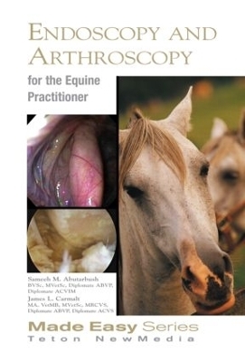
Equine Endoscopy and Arthroscopy for the Equine Practitioner - Sameeh Abutarbush, James Carmalt

Equine Endoscopy and Arthroscopy for the Equine Practitioner

Buch | Softcover
292 Seiten
2008
Teton NewMedia (Verlag)
978-1-59161-039-7 (ISBN)
75,95 inkl. MwSt
This addition to the Equine Made Series provides detailed, easy to read guidelines for performance of common endoscopic procedures in the horse. It includes step-by-step guidelines endoscopy of the upper airway, gastrointestinal, urinary and reproductive systems. A detailed section covering arthroscopy, including the temperomandicular joint, is especially useful to the equine practitioner. Normal anatomy is presented for orientation along with full coverage of abnormal conditions and their assessment. This fully illustrated text contains over 475 high quality clinical photographs and anatomic drawing which ease understanding.

Published by Teton New Media in the USA and distributed by CRC Press outside of North America.

Sameeh Abutarbush, James Carmalt

Contents

Section 1 Endoscopy Equipment, Use and Maintenance
General Principles 3
Some Helpful Hints 3

Section 2 Endoscopy of the Nasal Passages
Anatomy 12
Clinical Signs of Nasal Passage Disease 12
Endoscopic Technique 13
Diseases of the Nasal Passages 15
Rhinitis 15
Nasal Septum Deviation / Inflammation 16
Ethmoid Hematoma 17
Fungal Infection of the Ethmoid Labyrinth 18
Neoplastic Masses 18
Miscellaneous Diseases 19

Section 3 Endoscopy of the Paranasal Sinuses
Anatomy 22
Endoscopic Technique 25
Endoscopic Anatomy 26
Diseases of the Sinuses 27
Primary and Secondary Sinusitis 27
Neoplasia 28
Ethmoid Hematoma 28
Paranasal Sinus Cyst 28
Fractures and Trauma 29

Section 4 Endoscopy of the Guttural Pouch
Anatomy 34
The Medial Compartment 34
The Lateral Compartment 35
Endoscopic Technique 35
Entry into the Pouches (using a biopsy stylette) 36
Entry into the Pouches (using an artificial
insemination pipette) 37
Diseases of the Guttural Pouch 40
Tympany 40
Mycosis 42
Empyema 46
Temporohyoid Osteoarthropathy 49
Inflammation of the Guttural Pouch 51
Rupture of the Ventral Straight
Muscles of the Neck 52
Melanomatosis 52
Neoplasia 52
Cysts 53
Foreign Bodies 53

Section 5 Endoscopy of the Nasopharynx
Anatomy 56
Diseases of the Nasopharynx 56
Lymphoid Hyperplasia 56
Dorsal Displacement of the Soft Palate (DDSP) 58
Cleft Palate 60
Rostral Displacement of the Palatopharyngeal Arch 62
Collapse of the Dorsal Nasopharynx 62
Choanal Atresia 63
Nasopharyngeal Cicatrix 66
Nasopharyngeal Masses 67

Section 6 Endoscopy of the Larynx
Anatomy 70
Endoscopic Technique 71
Diseases of the Larynx 72
Aryepiglottic Fold Entrapment 72
Axial Deviation of the Aryepiglottic Folds 73
Epiglottic Retroversion 73
Epiglottitis 74
Subepiglottic Cysts 75
Arytenoid Chondritis 76
Left Laryngeal Hemiplegia 78
Right Laryngeal Hemiplegia 80
Biaxial Laryngeal Hemiplegia 80

Section 7 Endoscopy of the Trachea and Bronchi
Anatomy 84
Endoscopic Technique and Normal
Endoscopic Findings 84
Diseases of the Trachea 86
Tracheal Collapse 86
Tracheitis 86
Tracheal Luminal Obstruction and Masses 88
Miscellaneous Conditions 88
Gross Tracheal Discharge 88
Tracheal Wash and Aspirate 95
Bronchioalveolar Lavage 97

Section 8 Endoscopy of the Esophagus
Anatomy 102
Endoscopic Technique 103
Normal Endoscopic Findings 105
Diseases of the Esophagus 106
Esophageal Obstruction (Choke) 106
A-Primary choke 106
B-Secondary choke 113
Follow up 115
Treatment and Management 118
Esophagitis 121
Megaesophagus 122

Section 9 Endoscopy of the Stomach
Anatomy 126
Endoscopic Technique 127
Normal Endoscopic Findings 134
Diseases of the Stomach 135
Gastric Ulcers 135
Endoscopic Findings and Appearance of
Gastric Ulceration 137
Clinical Syndromes 139
Foals 139
Yearlings and Adults 139
Gastric Parasites 141
Gastric Neoplasia 143
Miscellaneous Gastric Conditions 143

Section 10 Endoscopy of the Duodenum
Anatomy 146
Endoscopic Technique 147
Normal Endoscopic Findings 148
Diseases of the Duodenum 151

Section 11 Endoscopy of the Urethra, Bladder and Ureters
Anatomy 154
Patient Preparation 155
Endoscopic Technique and
Normal Endoscopic Findings 155
Diseases of the Urinary System 162
Urolithiasis 162
Cystolithiasis 163
Sabulous Urolithiasis 165
Urethrolithiasis 166
Urinary Tract Infection 168
Urethritis 168
Cystitis 169
Pyelonephritis 170
Urethral Rent or Defect 171
Idiopathic Renal Hematuria 173
Exercise-Associated Hematuria 173
Urinary Tract Neoplasia 173
Ectopic Ureter 173
Urethrorectal Fistula 174

Section 12 Endoscopy of the Reproductive System of the Stallion
Normal Anatomic Features 178
Prepuce 178
Penis 180
Urethra 181
Pathologic Conditions 185
Urethritis 185
Summer Sores–Habronemiasis of the Urethra 185
Seminal Vesiculitis 186
Hemospermia 188
Congenital Anomalies 192

Section 13 Endoscopy of the Reproductive System of the Mare
Normal Anatomic Features 196
Endoscopic Technique 198
The Normal Endometrium 199
Endometrial Biopsy 203
Hysteroscopic Cannulation and Insemination 204
Pathologic Conditions of the Vagina 205
Urovagina 205
Vaginal Hematoma and Abscesses 206
Vaginal Lacerations 206
Varicose Veins 206
Vaginal Fusion 206
Pathologic Conditions of the Cervix 208
Cervical Lacerations 208
Cervical Adhesions 208
Pathologic Conditions of the Uterus 208
Endometritis 209
Endometrial, Glandular and Lymphatic Cysts 209
Pyometra 211
Persistent Endometrial Cups 212
Transluminal Adhesions 212
Tumors of the Uterus and Cervix 212
Foreign Bodies 214
Fetal Maceration or Mummification 215
Uterine Lacerations 215
Retained Fetal Membranes 215
Congenital Abnormalities 215
Pathologic Conditions of the Rectum 216
Rectal Stricture 216
Rectal Tears 216

Section 14 Arthroscopy
Introduction 220
Instrumentation 220
Pre- & Post-Surgical Considerations 222
Technique Hints 222
Common Pathological Conditions 223
Specific Joints (Approaches and Pathology) 224
Coffin Joint 224
Dorsal Approach 224
Palmar / Plantar Approach 226
Pastern Joint 229
Dorsal Approach 229
Palmar / Plantar Approach 230
Fetlock Joint 231
Dorsal Approach 231
Palmar / Plantar Approach 233
Carpal Joints (Radio- & Inter-Carpal
(Middle) Joints) 237
Radio-Carpal Joint 237
Inter-Carpal Joint 240
Elbow Joint 245
Craniolateral Approach 245
Caudoproximal Approach 247
Shoulder Joint (Scapulo-Humeral Joint) 248
Tibio-Tarsal Joint 252
Stifle Joint (Femoro-Patellar and
Femoro-Tibial Joint) 260
Femoro-Patellar 260
Femoro-Tibial
(Medial and Lateral Compartments) 265
Lateral Approach 265
Dorsal Approach 267
Hip Joint (Coxo-Femoral Joint) 268
Temporomandibular Joint 270
Tenoscopy and Bursoscopy 271
Navicular Bursa 271
Flexor Tendon Sheath 273
Extensor Tendon Sheaths 278
Carpal and Tarsal Sheath 278
Carpal Sheath 278
Tarsal Sheath 279
Bicipital Bursa 279

Index 281

Erscheint lt. Verlag 30.6.2008
Reihe/Serie Equine Made Easy Series
Verlagsort Jackson
Sprache englisch
Maße 146 x 215 mm
Gewicht 540 g
Themenwelt Naturwissenschaften Biologie Zoologie
Veterinärmedizin Pferd
Weitere Fachgebiete Land- / Forstwirtschaft / Fischerei
ISBN-10 1-59161-039-7 / 1591610397
ISBN-13 978-1-59161-039-7 / 9781591610397
Zustand Neuware
Haben Sie eine Frage zum Produkt?
Mehr entdecken
aus dem Bereich

von Rüdiger Wehner; Walter Jakob Gehring; Alfred Kühn

Buch | Softcover (2013)
Thieme (Verlag)
91,00