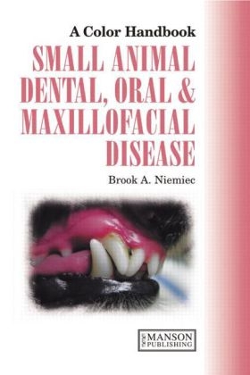
Small Animal Dental, Oral and Maxillofacial Disease
Manson Publishing Ltd (Verlag)
978-1-84076-108-5 (ISBN)
- Titel ist leider vergriffen;
keine Neuauflage - Artikel merken
In an area of growing interest to veterinarians, the authors have produced a rapid reference to the practical clinical aspects of small animal dentistry. The text is arranged to reflect the clinician’s thinking and approach to problems: background information, clinical relevance, key points, differential diagnoses, diagnostic tests, and management. Some 400 top-quality illustrations—color photos, imaging and diagrams—provide a critically important complement to the text.
The color handbook—here with revised text and references—offers real-life insights into the progression of oral disease and has been welcomed as a working resource by veterinary practitioners and students, and as a valuable review by more advanced veterinary dentists.
American Veterinary Dental College, Academy of Veterinary Dentistry, California Veterinary Specialties Group, California, USA
Anatomy and Physiology
Canine dental anatomy
Feline dental anatomy
Rodent and lagomorph dental anatomy
Dental terminology
Tooth development
Enamel, dentin, and pulp
Periodontium
Bones of the face and jaws
Muscles, cheeks, and lips
Neurovascular structures
Joints of the head
Hard and soft palates
Tongue
Salivary glands
Lymph nodes and tonsils
Oral Examination
Step 1: History
Step 2: General physical examination
Step 3: Orofacial examination
Step 4: Conscious (awake) intraoral examination
Step 5: The anesthetized orodental examination
Veterinary Dental Radiology
Step 1: Patient positioning
Step 2: Film placement within the patient’s mouth
Step 3: Positioning the beam head
Step 4: Setting the exposure
Step 5: Exposing the radiograph
Step 6: Developing the radiograph
Step 7: Techniques for various individual teeth
Step8 : Interpreting dental radiographs
Pathology in the Pediatric Patient
Persistent deciduous teeth
Fractured deciduous teeth
Malocclusions (general)
Deciduous malocclusions
Class I malocclusions
Mesioversed maxillary canines (lance effect)
Base narrow canines
Class II malocclusion (overshot, mandibular brachygnathism)
Class III malocclusion (undershot)
Class IV malocclusion (wry bite)
Cleft palate
Cleft lip (harelip)
Tight lip
Hypodontia/oligodontia and anodontia (congenitally
missing teeth)4
Impacted or embedded (unerupted) teeth
Dentigerous cyst (follicular cyst)
Odontoma
Hairy tongue
Enamel hypocalcification (hypoplasia)
Feline juvenile (puberty) gingivitis/periodontitis
Oral papillomatosis
Pathologies of the Dental Hard Tissues
Uncomplicated crown
fracture (closed crown fracture)
Complicated crown fracture (open crown fracture)
Caries (cavity, tooth decay)
Type feline tooth resorption (TR)
Type feline tooth resorption (TR)
Enamel hypoplasia and hypocalcification
Dental abrasion
Dental attrition
External resorption6
Internal resorption8
Intrinsic stains (endogenous stains)
Extrinsic stains (exogenous stains)
Primary endodontic lesion with secondary periodontal disease
Primary periodontal lesion with secondary endodontic involvement
Combined endodontic and periodontal lesion
Idiopathic root resorption
Problems with the Gingiva
Gingivitis
Periodontitis
Generalized gingival enlargement (gingival hyperplasia)
Trauma2
Epulids3
Gingivostomatitis (caudal stomatitis) in cats
Pathologies of the Oral Mucosa
Oronasal fistula
Eosinophilic granuloma complex
Chronic ulcerative paradental stomatitis (CUPS)
(kissing lesions)
Immune-mediated diseases affecting the oral cavity
Uremic stomatitis
Candidiasis (thrush)5
Caustic burns of the oral cavity
Problems with Muscles, Bones, and Joints
Masticatory myositis
Craniomandibular osteopathy
Idiopathic trigeminal neuritis
Temporomandibular joint luxation
Temporomandibular joint dysplasia
Fractures
Traumatic tooth avulsion and luxation
Root fractures
Osteomyelitis
Tumors and cysts
Hyperparathyroidism
Tetanus
Botulism
Malignant Oral Neoplasia
Introduction
Malignant melanoma
Fibrosarcoma
Squamous cell carcinoma
Histologically low-grade, biologically high-grade, fibrosarcoma
Osteosarcoma
Pathologies of the Salivary System
Sialoceles
Salivary gland tumors
Sialoliths (salivary stones)
Appendices
References
Index
| Erscheint lt. Verlag | 15.1.2010 |
|---|---|
| Reihe/Serie | Veterinary Color Handbook Series |
| Zusatzinfo | 401 Illustrations, color |
| Verlagsort | London |
| Sprache | englisch |
| Maße | 156 x 234 mm |
| Gewicht | 703 g |
| Einbandart | gebunden |
| Themenwelt | Veterinärmedizin ► Klinische Fächer ► Zahnheilkunde |
| Veterinärmedizin ► Kleintier ► Krankheitslehre | |
| ISBN-10 | 1-84076-108-3 / 1840761083 |
| ISBN-13 | 978-1-84076-108-5 / 9781840761085 |
| Zustand | Neuware |
| Haben Sie eine Frage zum Produkt? |
aus dem Bereich


