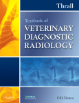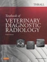
Textbook of Veterinary Diagnostic Radiology
Saunders (Verlag)
978-1-4160-2615-0 (ISBN)
- Titel erscheint in neuer Auflage
- Artikel merken
User-friendly and comprehensive, this essential resource covers all aspects of canine, feline, and equine diagnostic radiology and interpretation. It features relevant coverage of the physics of radiology, CT, and MRI, as well as valuable information on patient positioning and management, radiographic technique and safety measures, normal and abnormal anatomy, radiographic viewing and interpretation, and alternative imaging modalities. This edition features more than 500 additional images, a new chapter on the principles of digital imaging, and expanded coverage of brain and spinal cord imaging.
Section I: PHYSICS AND PRINCIPLES OF INTERPRETATION 1.Radiation Physics, Radiation Protection, and Darkroom Theory 2.Digital Images and Digital Radiographic Image Capture 3.Physics of Ultrasound Imaging 4.Physics of Computed Tomographic and Magnetic Resonance Imaging 5.Introduction to Radiographic Interpretation Section II: THE AXIAL SKELETON 6.Technical Issues and Interpretation Principles Relating to the Axial Skeleton 7.Normal CT, MR, and Radiographic Anatomy of the Axial Skeleton 8.The Canine Skull, Nasal Cavity, and Sinuses 9.Magnetic Resonance Imaging Features of Brain Disease 10.The Equine Skull, Nasal Cavity, and Sinuses 11.Canine Vertebrae 12.Canine Intervertebral Disk Disease Section III: THE APPENDICULAR SKELETON 13.Technical Issues and Interpretation Principles Relating to the Appendicular Skeleton 14.Normal Radiographic Anatomy of the Appendicular Skeleton 15.Orthopedic Diseases of Young and Growing Dogs and Cats 16.Fracture Healing and Complications 17.Canine and Feline Bone Tumors vs. Bone Infections 18.Canine and Feline Joint Disease 19.Equine Stifle and Tarsus 20.Equine Tarsus 21.Equine Metacarpus and Metatarsus 22.Equine Metacarpophalangeal and Metatarsophalangeal Joints 23.Equine Phalanges 24.Equine Navicular Bone Section IV: CARDIAC AND RESPIRATORY SYSTEMS 25.Technical Issues and Interpretation Principles Relating to the Cardiopulmonary System 26.Normal Radiographic Anatomy of the Cardiopulmonary System 27.The Canine and Feline Upper Airway and Trachea 28.The Canine and Feline Esophagus 29.The Canine and Feline Thoracic Wall 30.The Canine and Feline Diaphragm 31.The Canine Mediastinum 32.The Canine Pleural Space 33.The Canine and Feline Cardiovascular System 34.The Canine and Feline Lung 35.The Equine Lower Respiratory System Section V: CANINE AND FELINE ABDOMEN 36.Technical Issues and Interpretation Principles Relating to the Canine and Feline Abdomen 37.Normal Radiographic Anatomy of the Abdomen 38.The Peritoneal Space 39.The Liver and Spleen 40.The Kidney and Ureters 41.The Urinary Bladder 42.The Urethra 43.The Prostate 44.The Uterus, Ovaries, and Testes 45.The Stomach 46.The Small Bowel 47.The Large Bowel
| Zusatzinfo | Approx. 1500 illustrations |
|---|---|
| Verlagsort | Philadelphia |
| Sprache | englisch |
| Maße | 216 x 276 mm |
| Themenwelt | Veterinärmedizin ► Klinische Fächer ► Bildgebende Verfahren |
| ISBN-10 | 1-4160-2615-0 / 1416026150 |
| ISBN-13 | 978-1-4160-2615-0 / 9781416026150 |
| Zustand | Neuware |
| Informationen gemäß Produktsicherheitsverordnung (GPSR) | |
| Haben Sie eine Frage zum Produkt? |
aus dem Bereich


