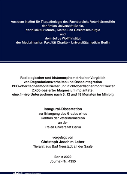
Radiologischer und histomorphometrischer Vergleich von Degradationsverhalten und Osseointegration PEO-oberflächenmodifizierter und nichtoberflächenmodifizierter ZX00-basierter Magnesiumimplantate
Eine in vivo Untersuchung nach 6, 12 und 18 Monaten im Minipig
Seiten
2023
|
1. Auflage
Mensch & Buch (Verlag)
978-3-96729-187-2 (ISBN)
Mensch & Buch (Verlag)
978-3-96729-187-2 (ISBN)
- Keine Verlagsinformationen verfügbar
- Artikel merken
Die chirurgische Frakturversorgung mit resorbierbaren Osteosynthesematerialien auf Magnesiumbasis hat sich in der Vergangenheit für manche Indikationen als vorteilhaft gegenüber nichtresorbierbarem Osteosynthesematerial wie Titan oder resorbierbaren Osteosynthesematerialien auf Polymerbasis erwiesen. Die Limitation für den routinemäßigen Einsatz ist das bei der Degradation frei werdende Wasserstoffgas, was in vorangegangenen Studien zu subkutanen und intraossären Gasansammlungen um das Implantat führte. Um einen stabilen Knochen-Implantat-Kontakt während der Frakturheilung zu gewährleisten und im späteren Verlauf der Degradation keine nachteiligen Effekte im Knochen um das Implantat zu erhalten, wurde mittels Legierung und Beschichtung von magnesiumbasierten Implantaten versucht, die Degradation zu verlangsamen. Ziel der Dissertation war es somit, erstmals im Großtierversuch das Degradationsverhalten sowie die Reaktion des umgebenden Knochens auf ZX00-Implantate mit und ohne PEO-Oberflächenmodifikation nach 6, 12 und 18 Monaten Implantationszeit zu untersuchen und gegenüberzustellen. Dafür wurden die Implantate in das Os frontale von Minipigs eingebracht und nach der jeweiligen Standzeit nach Explantation der Implantate mit umliegenden Knochen radiologisch und histomorphometrisch analysiert. Das Residualvolumen konnte in der Micro-CT-Untersuchung aufgrund der Dichte des Materials berechnet werden (SV/TV). In der histomorphometrischen Untersuchung wurde ebenfalls der noch nicht resorbierte Anteil an Implantationsmaterial mittels der Fläche des Implantatquerschnitts (SA/TA) berechnet. Die Ergebnisse beider Untersuchungsmethoden zeigen, dass das Residualvolumen der PEOoberflächenbeschichteten Implantate zu jedem Zeitpunkt höher ist und somit die Beschichtung erfolgreich die Degradation verlangsamt. Signifikante Unterschiede zeigten sich in der radiologischen Auswertung nach 12 und 18 Monaten Implantationszeit (p < 0,001), in der histomorphometrischen Untersuchung nach 12 Monaten (p < 0,001). Im Vergleich des Residualvolumens (SV/TV) zeigte sich nach 18 Monaten ein signifikant (p < 0,001) niedrigerer Wert als nach 6 Monaten bei den unbeschichteten Implantaten. Bei den beschichteten Implantaten war durch die verlangsamte Degradation der Unterschied nicht signifikant (p = 0,25).
Durch die verlangsamte Degradation der PEO-Oberflächenbeschichtung und der daraus resultierenden langsameren Freisetzung von Gas sollte die Bildung von Hohlräumen im Knochen reduziert und somit die Osseointegration verbessert werden. Radiologisch und histomorphometrisch wurden jeweils zwei Bereiche von der ursprünglichen Implantatgrenze bis 0,5 mm und bis 1,5 mm auf den Anteil an mineralisiertem Knochen innerhalb der Auswertungsbereiche (BV/TV in der radiologischen, BA/TA in der histomorphometrischen Untersuchung) untersucht. Die radiologischen Ergebnisse zeigen einen signifikant höheren BV/TV (p < 0,001) in beiden Bereichen nach 18 Monaten Implantationszeit bei den PEObeschichteten Implantaten. Histomorphometrisch ergaben sich die gleichen Tendenzen.
Im Vergleich des Volumenanteils an mineralisiertem Knochen (BV/TV) um die Implantate nach 6 und 18 Monaten Implantationszeit zeigten sich in beiden Untersuchungsbereichen die gleichen Tendenzen. Zwischen ursprünglicher Implantatgrenze und 0,5 mm Umgebung zeigt sich ein signifikant höherer mineralisierter Knochenanteil (p = 0,035) nach 18 Monaten beim beschichteten Implantat. Der BV/TV-Anteil nimmt hingegen bei den unbeschichteten Implantaten im Untersuchungsbereich bis 1,5 mm zur ursprünglichen Implantatgrenze im gleichen Untersuchungszeitraum signifikant ab (p = 0,034). Weshalb der Unterschied des Knochenvolumens erst zum letzten Untersuchungszeitpunkt auftritt, bleibt unklar. Ausgehend von den Ergebnissen dieser Studie wird vermutet, dass die PEO-Oberflächenbeschichtung im Untersuchungszeitaum bis 18 Monate post implantationem die Osseointegration durch verminderte Bildung von Hohlräumen verbessern kann. Radiological and histomorphometric comparison of degradation behavior and osseointegration of PEO-surface-modified and non-surface-modified ZX00-based magnesium implants: An in vivo study after 6,12 and 18 months in the Minipig
In the past the surgical treatment of fractures with resorbable magnesium-based osteosynthesis materials has been shown to be advantageous over non-resorbable osteosynthesis materials such as titanium or resorbable polymer-based osteosynthesis materials for some indications. The limitation for routine use is the hydrogen gas released during degradation, which led to subcutaneous and intraosseous gas accumulation around the implant in previous studies. To ensure stable bone-implant contact during fracture healing and to avoid adverse effects in the bone around the implant during the degradation process, alloying and coating of magnesium-based implants was used to try to slow down degradation. The aim of this doctoral thesis was thus to investigate the degradation behavior as well as the reaction of the surrounding bone to ZX00 implants with and without PEO surface modification in a large animal experiment after 6, 12 and 18 months of implantation. For this purpose, the implants were placed in the frontal bone of minipigs and analyzed radiologically and histomorphometrically after the respective standing time. The implants were explanted with the surrounding bone. The residual volume was calculated in the micro-CT examination based on the density of the material (SV/TV). In the histomorphometric examination, the proportion of the remaining implant material was calculated using the area of the implant’s cross-section (SA/TA). The results of the radiological and the histomorphometric evaluation show a higher residual volume of PEO surface-coated implants at all time points and prove the successful delay of magnesium corrosion. Significant differences were seen in the radiological evaluation after 12 and 18 months of implantation (p < 0.001), and in the histomorphometric examination after 12 months (p < 0.001). Comparison of residual volume (SV/TV) showed a significant (p < 0.001) lower value after 18 months than after 6 months for the uncoated implants. For the coated implants, due to the slowed degradation, the difference was not significant (p = 0.25).
The slowed degradation of the PEO surface coating and the resulting slower release of gas should reduce the formation of gas-associated lacunae and improves osseointegration. Radiologically and histomorphometrically, two areas each from the original implant margin up to 0.5 mm and up to 1.5 mm were examined for the proportion of mineralized bone within the evaluation areas (BV/TV in the radiological, BA/TA in the histomorphometric examination). The radiological results showed a significantly higher BV/TV (p < 0.001) in both areas after 18 months of implantation time in the PEO-coated implants. Histomorphometrically, the same trends emerged.
Comparison of the volume fraction of mineralized bone (BV/TV) around the implants after 6 and 18 months showed the same trends in both study areas. Between the original implant border and the 0.5 mm surrounding area, there is a significantly higher mineralized bone proportion (p = 0.035) after 18 months for the coated implant. In contrast, the BV/TV proportion decreases significantly in the uncoated implants in the study area up to 1.5 mm from the original implant boundary during the same study period (p = 0.034). Why the difference in bone volume density occurs only at the last examination time remains unclear. Based on the results of this study, it is suggested that the PEO surface coating may improve osseointegration by reducing the formation of gas-associated lacunae in the study period up to 18 months post-implantation.
| Erscheinungsdatum | 10.04.2024 |
|---|---|
| Verlagsort | Berlin |
| Sprache | deutsch |
| Maße | 148 x 210 mm |
| Gewicht | 320 g |
| Themenwelt | Veterinärmedizin ► Allgemein |
| Schlagworte | Bones • chirurgische Geräte • Experimental Surgery • Experimentelle Chirurgie • Fractures • Frakturen • Grafting • grafts • Knochen • Magnesium • miniature pigs • minipig • surgical equipment • Transplantate |
| ISBN-10 | 3-96729-187-1 / 3967291871 |
| ISBN-13 | 978-3-96729-187-2 / 9783967291872 |
| Zustand | Neuware |
| Informationen gemäß Produktsicherheitsverordnung (GPSR) | |
| Haben Sie eine Frage zum Produkt? |

