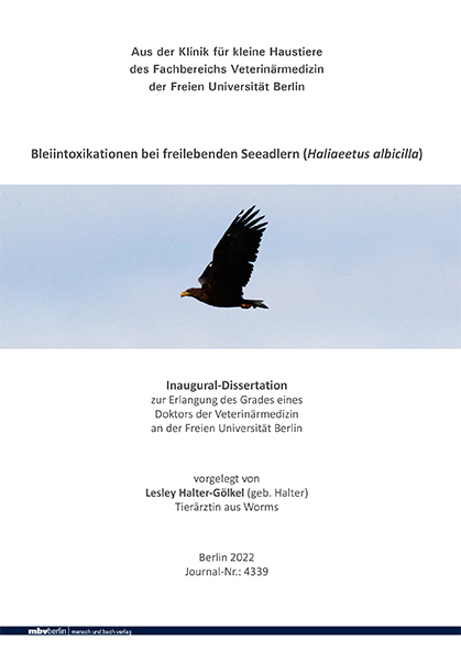
Bleiintoxikationen bei freilebenden Seeadlern (Haliaeetus albicilla)
Seiten
2022
|
1. Auflage
Mensch & Buch (Verlag)
978-3-96729-169-8 (ISBN)
Mensch & Buch (Verlag)
978-3-96729-169-8 (ISBN)
- Keine Verlagsinformationen verfügbar
- Artikel merken
Ziel dieser Studie war, medizinische Daten von freilebenden H. albicilla mit diagnostizierter Bleiintoxikation auszuwerten. Hierzu wurden klinische, bildgebende, blutchemische- und hämatologische Parameter sowie Blut- und Organbleikonzentrationen untersucht. Es wurde überprüft, ob das Vorhandensein verschiedener klinischer, bildgebender und labordiagnostischer Befunde Einfluss auf die Prognose einer Bleiintoxikation bei freilebenden H. albicilla hat. Über den Zeitraum von August 1998 bis Januar 2020 wurden Daten von 238 freilebenden, verletzt aufgefundenen H. albicilla, die in der Abteilung für Heim-, Zoo- und Wildtiere der Klinik für kleine Haustiere der Freien Universität Berlin vorgestellt wurden, ausgewertet. Als Einschlusskriterium für eine Bleiintoxikation wurde eine Blutbleikonzentration ab 0,4 ppm einhergehend mit typischen klinischen Symptomen gewählt. 31 % der Seeadler (73/238) wiesen eine Bleiintoxikation als alleinigen Befund auf und wurden in die Studie einbezogen. Die Seeadler stammten aus Brandenburg (44/73), Mecklenburg Vorpommern (14/73), Sachsen (6/73), Schleswig Holstein (4/73), Sachsen Anhalt (3/73) und Berlin (2/73). Ein Großteil der Seeadler wurde in den Herbst- und Wintermonaten (Oktober bis März) aufgefunden (60/73, 82 %). Die häufigste Altersgruppe waren adulte Seeadler (50/73, 69 %), gefolgt von immaturen (17/73, 23 %) und juvenilen Vögeln (4/73, 5 %) sowie Nestlingen (2/73, 3 %). Weibliche Seeadler (40/73, 55 %) wurden häufiger vorgestellt als männliche (30/73, 41 %). Typische klinische Untersuchungsbefunde, die bei 72 der 73 Seeadler erhoben werden konnten, waren Apathie (72/72, 100 %), Dyspnoe (50/72, 68 %), ein schlechter Ernährungszustand (44/72, 60 %), Biliverdinurie (35/72, 48 %), Erbrechen (23/72, 32 %), Kropfstase (21/72, 29 %) und neurologische Ausfallserscheinungen (19/72, 26 %). Bei der röntgenologischen Untersuchung konnten bei 44 % der Vögel metalldichte Fragmente im Verdauungstrakt nachgewiesen werden (32/73). Die Bleifragmente befanden sich am häufigsten im Magen, gefolgt von Darm und Kropf. Eine Hämolyse der Erythrozyten wurde bei 62 % (45/73) der Seeadler nachgewiesen. Die mediane Blutbleikonzentration lag bei 3,0 ppm (Min=0,4 ppm; Max=21,7 ppm). Signifikant höhere Blutbleikonzentrationen konnten bei Seeadlern mit Biliverdinurie (rs=0,44; p=0,002), Dyspnoe (rs=0,38; p=0,001) und metalldichten Fragmenten im Verdauungstrakt (rs=0,3; p=0,009) festgestellt werden. Zudem wiesen adulte Seeadler signifikant häufiger höhere Blutbleikonzentrationen auf als andere Altersgruppen (rs=0,25; p=0,03). Die hämatologische Untersuchung ergab bei 26 % der Seeadler (18/73) eine Anämie (Hämatokrit unter 29 %). Bei den blutchemischen Parametern konnten die stärksten Abweichungen von den Referenzintervallen bei der Aspartat Aminotransferase und der Kreatinkinaseaktivität sowie der Glukosekonzentration beobachtet werden. Eine therapeutische Endoskopie erfolgte bei 59 % (16/27) der Seeadler mit metalldichten Fragmenten im Magen. In 69 % (11/16) der Fälle wurden die Fragmente zwar erfolgreich entfernt, aber trotz Endoskopie verstarben 13 von 16 Seeadlern. Die Blutbleikonzentration konnte mit Ca Na2 EDTA 50 mg/kg KGW i.m. BID und 100 mg/kg KGW i.m. BID bzw. Ditripentat 50 mg/kg KGW i.m. SID intramuskulär kontinuierlich gesenkt werden. Insgesamt verstarben 79 % der Seeadler (31 weibliche, 25 männliche und zwei unbekannten Geschlechts) an den Folgen der Bleiintoxikation. Faktoren, die die Prognose signifikant beeinflussten, waren das Vorhandensein einer Dyspnoe (r=0,63; p=0,001), das Vorhandensein einer Biliverdinurie (r=0,43; p=0,005), die Höhe der Blutbleikonzentration (rs=0,36; p=0,002) sowie eine Hämolyse (r=0,31; p=0,01). Eine Überlebensbaumanalyse ergab, dass Dyspnoe und Anämie die einflussreichsten Faktoren für die Beurteilung der Prognose bei Seeadlern mit Bleiintoxikationen waren. Seeadler mit einer Dyspnoe wiesen eine Überlebenswahrscheinlichkeit von 6 % auf, Seeadler mit einer Anämie von 24 %.
Die Leberbleikonzentrationen der Seeadler mit Bleiintoxikation waren höher als die Nierenbleikonzentrationen (rs=0,8; p=0,001). 74 % (34/46) der verstorbenen Tiere wiesen deutlich erhöhte Leberbleikonzentrationen und 24 % (11/46) deutlich erhöhte Nierenbleikonzentrationen auf (>8 ppm). Bei 86 % der Seeadler, die mit einer Blutbleikonzentrationen unter 1 ppm verstarben (6/9), konnten Leber- und Nierenbleikonzentrationen von über 1 ppm nachgewiesen werden.
Dies ist die erste Studie, die den Verlauf einer Bleiintoxikation bei einer größeren Anzahl von Seeadlern auswertete. Die Ergebnisse dieser Arbeit können helfen, anhand klinischer, bildgebender und labordiagnostischer Befunde, die Prognose einer Bleiintoxikation bei Seeadlern besser zu beurteilen. Lead intoxication in free-ranging white-tailed eagles (Haliaeetus albicilla)
The aim of this study was to evaluate medical data from German white-tailed eagles with diagnosed lead poisoning based on clinical signs, diagnostic imaging and laboratory diagnostic findings. From August 1998 to January 2020 238 free-living H. albicilla were presented at the Small Animal Clinic of the Freie Universität Berlin. Inclusion criterion was a blood lead concentration of 0.4 ppm, typical clinical symptoms associated with lead intoxication and absence of any other clinical findings. Based on these, data of 73 free-living white-tailed eagles were included in this study. The majority of eagles originated from Brandenburg (44/73), followed by Mecklenburg Western Pomerania (14/73), Saxony (6/73), Schleswig Holstein (4/73), Saxony Anhalt (3/73) and Berlin (2/73). Most of the birds were found in autumn and winter months (October to March) (60/73, 82 %). The most common age group were adult white-tailed eagles (50/73, 69 %), followed by immature (17/73, 23 %) and juvenile birds (4/73, 5 %) as well as nestlings (2/73, 3 %). Female white-tailed eagles (40/73, 55 %) were presented more often than males (30/73, 41 %). Typical clinical findings could be evaluated in 72 out of 73 birds including apathy (72/72, 100 %), dyspnoea (50/72, 68 %), poor nutritional status (44/72, 60 %), biliverdinuria (35/72, 48 %), vomiting (23/72, 32 %), crop stasis (21/72, 29 %) and neurological signs (19/72, 26 %). Metal-dense fragments within the digestive tract were detected in 44 % of the birds during radiological examination (32/73). Those were most frequently located in the stomach, followed by intestines and crop. In 12 % of the white-tailed eagles, metal-dense fragments were furthermore found outside the digestive tract (4/34). Hemolysis was observed in the plasma of 62 % of all birds examined (45/73). The median blood lead concentration was 3.0 ppm (min=0.4 ppm; max=21.7 ppm). Significantly higher blood lead concentrations were found in white-tailed eagles presenting with biliverdinuria (rs=0.44; p=0.002), dyspnoea (rs=0.38; p=0.001), and metal dense fragments located within the digestive tract (rs=0.3; p=0.009). Furthermore, higher blood lead concentrations were significantly more common in adult white-tailed eagles (rs=0.25; p=0.03) than in other age groups. Hematological examination of white-tailed eagles revealed anemia in 26 % of the birds (18/73). The greatest deviations from reference intervals in blood biochemistry parameters were observed in aspartate aminotransferase and creatine kinase activity as well as in glucose concentration. Therapeutic endoscopy was attempted in 59 % (16/27) of white-tailed eagles with metal-dense fragments located in the stomach. Lead fragments were successfully retrieved in 69 % (11/16) of birds, but despite endoscopic treatment 13 of 16 white-tailed eagles died due to lead intoxication. Blood lead concentration could be continuously reduced with intramuscular administration of Ca Na2 EDTA at 50 mg/kg BW BID, at 100 mg/kg BW BID or Ditripentate at 50 mg/kg BW SID. Overall, 79 % of the white-tailed eagles (31 females, 25 males and two unknown sex) presented died as a result of lead poisoning. Factors that significantly influenced the prognosis were the presence of dyspnoea (r=0.63; p=0.001), biliverdinuria (r=0.43; p=0.005), blood lead concentration (rs=0.36; p=0.002) as well as hemolysis (r=0.31; p=0.01). A survival tree analysis indicated that dyspnea was the most relevant factor for assessing the prognosis of white-tailed eagles with lead intoxication followed by the presence of anemia. White-tailed eagles with dypnoea had a 6 % survival rate, while the chance of survival of birds with anemia was 24 %.
Postmortem liver lead concentrations of white-tailed eagles were higher than kidney lead concentrations (rs=0.8; p=0.001). 74 % (34/46) of the birds showed significantly increased liver lead concentrations while 24 % (11/46) had increased kidney lead concentrations (>8 ppm). In 86 % of the white-tailed eagles dying with blood lead concentrations of less than 1 ppm (6/9), liver lead concentrations higher than 1 ppm were detected.
To the author’s knowledge this is the first study evaluating the course of lead intoxication in a larger number of white-tailed eagles. The results of this study should therefore help to better assess the prognosis of lead intoxication in this species and treat affected birds accordingly.
| Erscheinungsdatum | 09.04.2024 |
|---|---|
| Verlagsort | Berlin |
| Sprache | deutsch |
| Maße | 148 x 210 mm |
| Gewicht | 300 g |
| Themenwelt | Veterinärmedizin ► Kleintier |
| Schlagworte | Anaemia • clinical examination • Eagles • lead • lead poisoning • Liver • predatory birds • Symptoms • Toxicity |
| ISBN-10 | 3-96729-169-3 / 3967291693 |
| ISBN-13 | 978-3-96729-169-8 / 9783967291698 |
| Zustand | Neuware |
| Informationen gemäß Produktsicherheitsverordnung (GPSR) | |
| Haben Sie eine Frage zum Produkt? |
Mehr entdecken
aus dem Bereich
aus dem Bereich
Clinical Medicine and Management
Buch | Hardcover (2024)
Elsevier - Health Sciences Division (Verlag)
199,95 €
A Book of Lists
Buch | Softcover (2024)
Saunders (Verlag)
59,95 €
