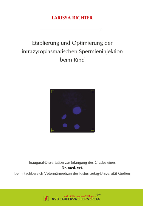
Etablierung und Optimierung der intrazytoplasmatischen Spermieninjektion beim Rind
Seiten
2022
VVB Laufersweiler Verlag
978-3-8359-7040-3 (ISBN)
VVB Laufersweiler Verlag
978-3-8359-7040-3 (ISBN)
- Keine Verlagsinformationen verfügbar
- Artikel merken
Die intrazytoplasmatische Spermieninjektion (ICSI) beim Rind benötigt zur Entwicklung der Embryonen ein geeignetes Aktivierungsverfahren. Die Problematik besteht jedoch beim Rind darin, dass so auch parthenogenetische entstehen, welche morphologisch nicht von tatsächlich befruchteten Stadien zu unterscheiden sind. Ziel dieser Arbeit war es, eine paternale Beteiligung nach der ICSI nachzuweisen und somit die tatsächliche Befruchtung. Dazu mussten die parthenogenetischen Entwicklungsstadien von den tatsächlich befruchteten unterschieden werden. Für eine erste Einschätzung wurden die Zygoten 18 Stunden nach der initialen Aktivierung mit Hoechst 33342 oder mit Hoechst und dem MitoTracker Green FM® gefärbt. Der MitoTracker Green FM® ermöglichte eine Zuordnung des Spermienmittelstücks und identifizierte somit den paternalen Vorkern. Die Ergebnisse wurden sowohl für die ICSI-Gruppe als auch für die Kontrollgruppen bestehend aus einer In-vitro-Fertilisations- (IVF), Chemisch-aktivierten- (CA), CA und Sham-injizierten- (Sham) Gruppe verglichen. Die erfolgreiche haploide Aktivierung wurde durch das Ausschleusen des zweiten Polköpers bestätigt. Zudem erfolgte ein Vergleich der Ergebnisse der Embryonen der diploid und der haploid aktivierten Gruppen, sowohl mit denen, die mit gesextem als auch ungesextem Sperma erstellt wurden. Die Erhebung der Teilungs- und Entwicklungsraten für alle Gruppen und deren Vergleich ließen somit Rückschlüsse auf den Anteil an parthenogenetischen Entwicklungsstadien zu. Zusätzlich erfolgten Versuche mit Y-chromosomal-gesextem Sperma durchgeführt. An Tag sieben oder acht wurden Embryonen für die RT-qPCR aus den Versuchen mit gesextem und ungesextem Sperma eingefroren. Auf molekularer Ebene fand die Erfassung der Transkriptmengen von sieben entwicklungsrelevanten Genen (DNMT1, DNMT3a, IGF2R, G6PD, NNAT, BAX und BCL2L1) statt. An einer repräsentativen Stichprobe an Embryonen wurde das Geschlecht der Embryonen durch eine PCR ermittelt, um somit einen Rückschluss auf den Anteil parthenogenetischer Entwicklungsstadien zu erhalten. Bei Embryonen der ICSI-Gruppe, welche mit Y-chromosomal-gesextem Sperma erzeugt wurden, stellt der Anteil an männlichen Embryonen den Anteil der tatsächlich befruchteten Embryonen dar. Die Durchführung einer RT-qPCR, die die relative Transkriptmenge eines paternales Gentranskriptes wie bspw. NNAT ermittelte und lieferte somit einen Nachweis der paternale Beteiligung. Zum anderen erfolgte der Vergleich der Qualität der Embryonen der CA, Sham-, ICSI- und IVF-Gruppe über die Untersuchung unterschiedlicher Transkripte wie bspw. IGF2R, BCL2L1 und BAX als Qualitätsmarker.
Es konnten folgende Ergebnisse erzielt werden:
1.Färbung mit Hoechst 33343:
a.In der ICSI-Gruppe waren 26,2 % normal und erfolgreich befruchtet (2PN/2PB)
b.In der IVF-Gruppe waren 60,5 % normal und erfolgreich befruchtet (2PN/2PB)
c.In der CA-Gruppe waren 78,4 % korrekt aktiviert (2PB und 1PN)
d.In der Sham-Gruppe mit Aktivierung waren 24,8 % korrekt aktiviert (2PB und 1PN)
2.Färbung mit Hoechst 33343 und MitoTracker Green FM®:
a.In der ICSI-Gruppe waren 45,5 % normal und erfolgreich befruchtet (2PB+2PN+Mittelstück des Spermiums)
b.In der IVF-Gruppe waren 50,5 % normal und erfolgreich befruchtet (2PB+2PN+ Mittelstück des Spermiums)
3.Teilungsraten:
a.Bei den Embryonen der diploiden Gruppe waren signifikante Unterschiede zwischen denen der ICSI- (53,7 %) und denen der IVF- (70,5 %) und denen der Sham-Gruppe mit chemischer Aktivierung (44,3 %) feststellbar.
b.Bei den Embryonen der haploid aktivierten Gruppe lagen signifikante Unterschiede zwischen denen der ICSI (41,9 %) und der Sham-Gruppe (4,5 %), sowie zu der IVF-Gruppe (67,7 %) vor.
c.Der Vergleich der Embryonen der haploid aktivierten und der diploid aktivierten Gruppen ergab signifikante Unterschiede zwischen den beiden ICSI-Gruppen (diploid: 53,7 %; haploid: 41,9 %)
d.Bei den Embryonen der ICSI-Gruppe mit haploider Aktivierung und denen mit gesextem Sperma waren signifikante Unterschiede zwischen X-chromosomal gesextem Sperma (51,2 %) und Y-chromosomal gesextem Sperma des Bullen zwei (40,9 %) vorhanden. Zudem lagen signifikante Unterschiede zwischen Y-gesextem Sperma des Bullen zwei (40,9 %) und drei (28,5 %) vor.
4.Entwicklungsraten:
a.In der diploid aktivierten Gruppe wiesen die Embryonen der CA-Gruppe (18,7 %) im Vergleich zu denen der ICSI-Gruppe keine große Abweichung auf (24,1 %), somit enthält die ICSI-Gruppe wahrscheinlich einen hohen Anteil parthenogenetischer Entwicklungsstadien.
b.Die reine Sham-Injektion führte weder zu Teilungs- noch zu Entwicklungsraten, es fand also keinerlei Entwicklung der Embryonen statt.
c.Bei den Embryonen der haploid aktivierten Gruppe wiesen die der IVF-Gruppe (31,7 %) signifikante Unterschiede gegenüber denen der CA- (9,2 %) und der ICSI-Gruppe (14,8 %) auf.
d.Der Vergleich der Embryonen der haploid und der diploid aktivierten Gruppen ergab signifikante Unterschiede zwischen denen der IVF-Gruppe und beiden CA-Gruppen (IVF: 31,7 %; diploid: 18,7 %; haploid: 9,2 %)
e.In Embryonen der Gruppe mit gesextem Sperma wiesen 90,9 % der Embryonen an Tag sieben das Stadium der Morula auf, wovon sich nur 7,1 % bis zum nächsten Tag weiter zur Blastozyste entwickelten
5.Sexing
Die Embryonen der CA-Gruppen, die der X-chromosomal-gesexten Gruppe und die der Sham-Gruppe mit diploider Aktivierung zeigten mit 100 % weiblichen Embryonen das erwartete Geschlechtsverhältnis. Ebenso erfüllten die Embryonen der IVF-Gruppen mit 33,3 % Anteil an weiblichen Embryonen bei den Morulae und 67,9 % bei den Blastozysten die Erwartungen zur Geschlechtsverteilung. Für Stadien der ICSI-Gruppe mit diploider Aktivierung wurde zumindest ein Teil an männlichen Embryonen erwartet, dieser lag jedoch bei 0 %. Für die der haploid aktivierten Gruppe wurde eine ca. 50:50 Aufteilung erwartet, der Anteil weiblicher Embryonen lag jedoch bei 93,3 %. Für beide ICSI-Entwicklungsstadien (mit Y-chromosomal gesextem Sperma erstellt) wurde eine 100 % männliche Verteilung erwartet. Sie lag jedoch bei den Blastozysten bei 100 % und bei den Morulae bei 63,6 % weiblichen Embryonen
6.Transkripte:
a.DNMT1: Eine signifikant erhöhte Transkriptmenge wurde in den Embryonen der diploid aktivierten ICSI-Gruppe und der IVF-Gruppe gegenüber den Embryonen der diploiden CA-Gruppe und der haploid aktivierten Gruppe festgestellt.
b.DNMT3a: In den Embryonen der diploid aktivierten ICSI-Gruppe lagen signifikant erhöhte Transkriptmengen gegenüber allen anderen Embryonen aller Gruppen vor. Ebenso wurde eine Erhöhung bei den Blastozysten der IVF-Gruppe gegenüber der bei den Morulae in der IVF-Gruppe und der ICSI-Gruppe mit Y-chromosomal gesextem Sperma detektiert.
c.IGF2R: Es lag eine signifikant erhöhte Transkriptmenge in den Blastozysten der diploid aktivierten ICSI-Gruppe gegenüber denen der haploid aktvierten Morula der ICSI-Gruppe mit Y-chromosomal gesextem Sperma vor. Ebenso wiesen die Stadien der IVF-Gruppe bei den Blastozysten eine signifikant erhöhte Transkriptmenge gegenüber den Morulae aus der Y-chromosomal gesexten ICSI-Gruppe auf.
d.NNAT: Es konnte eine tendenziell höhere Transkriptmenge für die Blastozysten der IVF-Gruppe gegenüber den Embryonen aller anderen Gruppen ermittelt werden. Ebenso war die Transkriptmenge in den weiblichen Blastozysten der IVF-Gruppe gegenüber denen der Morula signifikant erhöht.
e.G6PD: Die Transkriptmenge der Embryonen der ICSI-Gruppe mit diploider Aktivierung war signifikant gegenüber denen aller anderen Gruppen erhöht, ausgenommen bei der IVF-Gruppe mit weiblichen Blastozysten. Ebenso war ein signifikanter Unterschied zwischen den weiblichen und männlichen Blastozysten der ICSI-Gruppe und den männlichen Morulae der ICSI-Gruppe zu erkennen.
f.BCL2L1: Die Embryonen der diploiden ICSI-Gruppe wies eine signifikant höhere Transkriptmenge als die der beiden CA-Gruppen und die der Gruppe der Morula aus der Gruppe mit Y-chromosomal gesextem Sperma auf. Die Transkriptmenge der Blastozysten der IVF-Gruppe war signifikant gegenüber der der weiblichen Morulae der ICSI-Gruppe mit Y-chromosomal-gesextem Sperma erhöht.
g.BAX: Die relative Transkriptmenge der Embryonen in der ICSI-Gruppe mit diploider Aktivierung war signifikant gegenüber denen der weiblichen Morula der ICSI-Gruppe mit Y-chromosomal-gesextem Sperma erhöht. Ebenso wiesen die Blastozysten der IVF-Gruppe signifikant höhere Werte als die Morulae der ICSI-Gruppe auf.
Abschließend kann zusammengefasst werden, dass das Sexing der ICSI-Gruppe mit Y-chromosomal gesextem Sperma konkrete Rückschlüsse auf den Anteil parthenogenetischer Embryonen zulässt. Dieser Anteil ist sowohl im Morulae- als auch Blastozystenstadium sehr hoch. Die anderen Nachweisverfahren und Gruppen lassen nur Tendenzen für einen hohen Anteil parthenogenetischer Embryonen erkennen. Weitere geeignete Methoden müssen erforscht und etabliert werden um eindeutige Aussagen für ICSI-Gruppen nach Verwendung ungesextem Spermas zu erlangen. Der Anteil tatsächlich befruchteter Stadien scheint somit niedrig zu sein. Um die ICSI als konventionelles Verfahren der IVP zu etablieren ist es somit von großer Bedeutung im ersten Schritt parthenogenetische Stadien zu erfassen und nur tatsächlich befruchtete Stadien zu übertragen. Daran anschließend muss es zu einer Verbesserung der Aktivierungsverfahren hin zu einem höheren Anteil tatsächlich befruchteter Stadien kommen. Zum einen wäre es sinnvoll Aktivierungsprotokolle zu verwenden, die zu einem hohen Anteil eine haploide Aktivierung der Eizelle verursachen. Durch den Einsatz einer Vorbehandlung der Spermien und einem geeigneten Aktivierungsprotokoll nach der ICSI wäre zudem eine bessere paternale Vorkernausbildung möglich. In bovine ICSI a suitable activation procedure is necessary for embryonic development. In this study, an activation protocol with ionomycin and 6-DMAP was used. The problem to solve is, that all these activation methods can also cause a parthenogenetic development of the oocytes. Morphologically these stages cannot be distinguished from correct fertilized stages. The aim of this study was to check the paternal involvement after ICSI and thus the correct fertilization. To do this, the parthenogenetic stages of development had to be distinguished from those that were correct fertilized. For an initial assessment, the zygotes were stained 18 hours after the activation with Hoechst 33342 or with Hoechst and the MitoTracker Green FM®. The MitoTracker Green FM® stained the mitochondria in the midpiece of the sperm and thus identified the paternal pronucleus. The results were compared for embryos of both, the ICSI group and the control groups consisting of an IVF, CA, CA and Sham group. Successful haploid activation was confirmed by extrusion of the second polar body. In addition, the results of the embryos of the diploid and haploid activated groups were compared with those with sexed and unsexed sperm. The cleavage and development rates were counted and compared within all embryo groups in order to draw conclusions about the proportion of parthenogenetic developmental stages. In addition, ICSI experiments were carried out with Y-chromosomal sexed sperm. At day 7 or 8, embryos were frozen for RT-qPCR analyses from the sexed and unsexed sperm experiments. To analyze the molecular level the transcript amounts of seven developmentally relevant genes (DNMT1, DNMT3a, IGF2R, G6PD, NNAT, BAX and BCL2L1) were determined. In addition, the RT-qPCR for NNAT was carried out, which determines a paternal gene transcript which should prove the paternal involvement. The gender of the embryos was determined by PCR on a representative sample of embryos in order to draw conclusions about the proportion of parthenogenetic stages of development. In the ICSI embryo group, that were created with Y-chromosomal sexed sperm, the proportion of male embryos represents the proportion of embryos correct fertilized.
The following results could be achieved:
1. Staining with Hoechst 33343:
a.In the ICSI group, 26.2% were fertilized normally and successfully (2PN/2PB).
b.In the IVF group, 60.5% were normally and successfully (2PN/2PB) fertilized.
c.In the CA group, 78.4 % were correctly activated (2PB/1PN).
d.In the Sham Group, 24.8 % were correctly activated (2PB/1PN).
2.Staining Hoechst 33343 and MitoTracker Green FM®:
a.In the ICSI-group, 45.5 % were normally and successfully fertilized (2PB/2PN/middle piece of the sperm).
b.In the IVF-group, 50.5 % were normally and successfully fertilized (2PB/2PN/middle piece of the sperm).
3.Cleavage rates:
a.Embryos of the diploid group, showed significant differences between those of the ICSI group (53.7 %), of the IVF group (70.5 %) and of the Sham group with chemical activation (44.3 %).
b.For embryos of the haploid group, significant differences were detected between those of the ICSI group (41.9 %), the IVF group (67.7 %) and the Sham-group (4.5 %).
c.Comparing embryos after haploid and diploid activation differences were dertermined between those of the ICSI groups (diploid 53.7 %, haploid 41,9 %).
For embryos of the haploid ICSI group generated with sex sorted sperm significant differences were realized between those produced with X (51,2 %) and Y sorted sperm from bull two (40.9 %). There were also significant differences between embryos stemming from the Y sorted sperm from bull two (40.9 %) and bull three (28.5%)
4.Developmental rates :
a.The embryos of the diploid activated CA group (18.7 %) did not show any deviations compared to those of the ICSI group (24.1 %). The ICSI group probably contains a high proportion of parthenogenetic stages.
b.Sham injection without activation resulted in no development.
c.The embryos of the haploid activated group significantly differed between those of the CA group (9.2 %), the ICSI group (14.8 %) and the IVF group (31.7 %).
d.The comparison of the embryos between the haploid and the diploid activated groups revealed significant differences between those of the IVF group and both CA groups (IVF: 31.7 %; diploid: 18.7 %; haploid: 9.2 %).
e.In the group of embryos generated with sexed sperm, 90.9% of the embryos showed the morula stage on day 7, of which only 7.1% developed into a blastocyst by the next day.
5.Sexing
Embryos of the CA groups, the X linked sexed group and the sham group with diploid activation showed the expected sex ratio with 100% female ones. The embryos of the IVF groups also met the expectations for gender distribution with 33.3% proportion of female ones at the morula and 67.9% at the blastocyst stage. For embryos of the ICSI group with diploid activation, at least some male embryos were expected, but there were 0%. For embryos the haploid activated group an approx. 50:50 distribution was expected, but the proportion of female embryos was 93.3 %. A 100% male allocation was expected for both Y-chromosomally sexed ICSI developmental stages. However, it was 100% female at the blastocyst stage and 63.6% for morulae.
6.Transcripts:
1.DNMT1: A significantly increased amount was detected in embryos of the diploid activated ICSI group and the IVF group compared to the embryos of the diploid CA group and the haploid activated group.
2.DNMT3a: Embryos of the diploid activated ICSI group showed significant higher amounts compared to those of all other groups. An increase was also detected in the blastocysts of the IVF group compared to the morula of the IVF group and the ICSI group generated with Y chromosomal sexed sperm.
3.IGF2R: There was a significant increased amount of transcript in the embryos of the diploid activated ICSI group compared those of the haploid activated morula of the ICSI group generated with Y linked sexed sperm. Blastocysts of the IVF group also showed a significantly increased amount of transcripts compared to the morula from the Y chromosomal sexed ICSI group.
4.NNAT: It was possible to determine a tendency towards a higher amount of transcripts for blastocysts of the IVF group compared the ones of all other groups. The amount of transcripts in female blastocysts of the IVF group was also significantly higher than that of the morula.
5.G6PD: The number of transcripts in embryos of the ICSI group generated with diploid activation was significantly higher than that of all embryos from the other groups, except for those of the IVF group with female blastocysts. There was also a significant difference between female and male blastocysts of the ICSI group and the male morula of the ICSI group.
6.BCL2L1: The embryos of the diploid ICSI group showed a significantly higher amount of transcript than those of the two CA groups and the group of morulae from the group with Y chromosomal sexed sperm. The transcript amount of blastocysts from IVF group was significantly increased compared to that of the female morulae of the ICSI group produced with Y chromosome sorted sperm.
7.BAX: The relative amount of transcripts for embryos in the ICSI group with diploid activation was significantly higher than that of female morula of the ICSI group with Y sorted sperm. Blastocysts of the IVF group had also significantly higher values than the morulae of the ICSI-group.
In conclusion, it can be summarized that the sexing of the ICSI group with Y chromosome sexed sperm allows specific conclusions to be drawn about the proportion of parthenogenetic embryos. This proportion is very high in both, the morulae and blastocyst stage. The other detection methods and groups only show tendencies for a higher proportion of parthenogenetic embryos. Further methods must be tested and established in order to obtain unambiguous statements regarding the sex if embryos generated via ICSI with non-sexed Sperm. The proportion of correct fertilized stages thus seems to be low. In order to establish ICSI as a conventional method of IVP, it is therefore of great importance to record parthenogenetic stages in a first step and to only transfer correct fertilized stages. This must be followed by an improvement in the activation process towards a higher proportion of correct fertilized stage.
| Erscheinungsdatum | 27.06.2022 |
|---|---|
| Reihe/Serie | Edition Scientifique |
| Verlagsort | Gießen |
| Sprache | deutsch |
| Maße | 210 x 148 mm |
| Gewicht | 250 g |
| Themenwelt | Veterinärmedizin ► Allgemein |
| Veterinärmedizin ► Großtier ► Rind | |
| Schlagworte | Befruchtung • Spermien • Vermehrung |
| ISBN-10 | 3-8359-7040-2 / 3835970402 |
| ISBN-13 | 978-3-8359-7040-3 / 9783835970403 |
| Zustand | Neuware |
| Informationen gemäß Produktsicherheitsverordnung (GPSR) | |
| Haben Sie eine Frage zum Produkt? |
Mehr entdecken
aus dem Bereich
aus dem Bereich
Buch | Hardcover (2021)
Schlütersche (Verlag)
159,00 €


