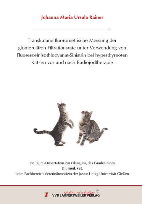
Transkutane fluorometrische Messung der glomerulären Filtrationsrate unter Verwendung von Fluoresceinisothiocyanat-Sinistrin bei hyperthyreoten Katzen vor und nach Radiojodtherapie
Seiten
2021
VVB Laufersweiler Verlag
978-3-8359-6955-1 (ISBN)
VVB Laufersweiler Verlag
978-3-8359-6955-1 (ISBN)
- Keine Verlagsinformationen verfügbar
- Artikel merken
Chronische Nierenerkrankungen und die Hyperthyreose sind häufige Erkrankungen der geriatrischen Katze, die sich gegenseitig beeinflussen. Vorhandene Blut- und Urin-Parameter der Routine-Diagnostik sind wenig sensitiv um Nierenerkrankungen frühzeitig zu erkennen und der beste Parameter, um die Nierenfunktion widerzuspiegeln, ist die Glomeruläre Filtrationsrate (GFR). Die vorhandenen Clearance-Messmethoden zur Bestimmung der GFR sind arbeits- und zeitaufwendig, belastend für die Patienten sowie mitunter kostenintensiv und infolgedessen wenig geeignet für den Einsatz im klinischen Alltag. Nach neuen, einfachen und kostengünstigen Messverfahren zur Bestimmung der GFR wird fortwährend gesucht und eine relativ neue Messmethode ist die transkutane, fluorometrische Messung von FITC-Sinistrin (FITC-S) mit einem Miniatur-Fluoreszenzmessgerät (NIC-Kidney-Device, Mannheim Pharma & Diagnostics GmbH). Die Messmethode wurde bereits erfolgreich an Kleinnagern in der medizinischen Forschung eingesetzt und es gibt erste erfolgversprechende Untersuchungen an gesunden Hunden und Katzen. Es ist bekannt, dass eine Hyperthyreose zu einer gesteigerten glomerulären Filtratonsrate führt und die erfolgreiche Therapie einer Schilddrüsenüberfunktion mit einem Sinken der GFR einhergeht. Infolgedessen stellt die Untersuchung von hyperthyreoten Katzen vor sowie nach RJT ein gutes Modell zur Überprüfung von GFR-Messmethoden zur Detektion von Veränderungen der Nierenfunktion dar.
Ziel der vorliegenden Studie war, die Anwendbarkeit des transkutanen fluorometrischen Messsystems an hyperthyreoten Katzen vor und nach RJT im Vergleich zu einer etablierten, invasiven Sinistrin-Plasma-Clearance zu überprüfen. Weiterhin sollten das 1- und 3-Kompartimentmodell als mögliche pharmakokinetische Modelle zur Beschreibung der transkutanen Eliminationskurven erprobt werden und die im Vorhinein ermittelte Dosierung von 30mg/kg FITC-S sowie die Messlokalisation am ventralen Abdomen von Katzen überprüft werden.
In die prospektive Studie wurden acht Katzen mit einer Hyperthyreose eingeschlossen, die zur Radiojodtherapie (RJT) in der Abteilung für Innere Medizin der Klinik für Kleintiere der Justus-Liebig-Universität Gießen vorgestellt worden sind. Es wurden die regelmäßigen Voruntersuchungen vor der RJT bestehend aus klinischer Untersuchung, Hämatologie, Blutchemie, einer Urinuntersuchung, kardiologischen Untersuchung inklusive Echokardiographie, Röntgen des Thorax und einer Blutdruckmessung sowie einer basalen T4-Konzentration durchgeführt. Die Routine-Kontrolluntersuchung bestehend aus T4-Messung, Blutbild, Blutchemie und Urinuntersuchung erfolgte 1 Woche nach der RJT. Zusätzlich wurden vor der RJT sowie 2 Wochen danach parallel eine transkutane, fluorometrische Messung einer FITC-Sinistrin-Clearance und eine invasive Sinistrin-Plasma-Clearance aus seriell entnommenen Blutproben durchgeführt. Die etablierte Sinistrin-Plasma-Clearance diente hierbei als Referenzmethode und die transkutane Messung erfolgte zeitgleich mittels 2 Miniaturfluoreszenzmessgeräten, die am ventralen kranialen sowie kaudalen Abdomen der Katzen platziert worden sind. Die Berechnung der Plasma-Clearance von Sinistrin (GFRp) sowie der Halbwertszeit (HWZp) erfolgte mit einem etablierten 2-Kompartimentmodell und die Kalkulation der transkutanen Halbwertszeiten mit einem 1-Kompartimentmodell (HWZtk1e) sowie 3-Kompartimentmodell (HWZtk3e). Angelehnt an vorangegangene Studien zur transkutanen Messmethde an Kleinnagern wurde anschließend die transkutane FITC-S-Clearance (GFRtk1e/GFRtk3e) über einen semiempirischen, speziesspezifischen Konversionsfaktor berechnet. Alle Parameter wurden auf signifikante Unterschiede zu den beiden Untersuchungszeitpunkten vor und nach RJT untersucht. Mittels linearer Regression wurde die Korrelation zwischen HWZp und HWZtk1e sowie HWZtk3e und zwischen GFRp und GFRtk1e sowie GFRtk3e untersucht.
3/8 Katzen waren zum Zeitpunkt der Kontrolluntersuchung nach RJT noch hyperthyreot. Von 6 Katzen lagen zusätzlich T4-Werte 3 Monate nach RJT vor und zu diesem Zeitpunkt waren 5/6 Katzen euthyreot und 1 Katze hatte labordiagnostisch ein erniedrigtes T4 ohne klinische Hinweise auf eine Hypothyreose. Die Resultate der Routine-Untersuchungen vor und nach RJT stimmten mit den vorhandenen Literaturangaben überein.
Die invasive Messung der Sinistrin-Elimination ergab einen signifikanten Unterschied von HWZp (p= 0,0328) und GFRp (p= 0,0078) zwischen den Messzeitpunkten vor und nach RJT. Die GFRp zeigte bei allen Katzen unabhängig von der Thyroxinkonzentration nach RJT eine Verminderung der Nierenfunktion.
Die transkutane, fluorometrische Messung der FITC-S-Elimination war einfach durchzuführen, verlief komplikationslos und wurde von allen Studienpatienten gut akzeptiert. Die transkutanen Eliminationskurven zeigten hochgradige Kurvensprünge infolge von Bewegungsartefakten, allerdings waren die Bewegungsartefakte bei der Messung am kaudalen Abdomen im Vergleich zur kranialen Messposition schwerwiegender. Unter Berücksichtigung der gleichmäßigen Eliminationskurven in vorangegangenen Studien an Hunden und Kleinnagern, mit einer Messlokalisation an der seitlichen Brustwand sowie auf dem Rücken, muss die Messposition am ventralen Abdomen bei Katzen als inadäquat bewertet werden. 13/32 der ermittelten transkutanen Eliminationskurven zeigten ein niedriges maximales Fluoreszenzsignal, das für eine unzureichende Dosierung des FITC-Sinistrins spricht. Demnach war bei 6/8 Katzen die Dosierung von 30mg/kg zu niedrig und kann nicht als allgemeingültige Dosierungen für die transkutane GFR-Messung bei Katzen bestätigt werden. Für alle ermittelten transkutanen Eliminationskurven konnte eine Halbwertszeit mittels 1- und 3-Kompartimentmodell bestimmt werden, allerdings müssen die Ergebnisse sowie ihre Validität unter Berücksichtigung der niedrigen Qualität der Eliminationskurven kritisch hinterfragt werden. Die mittels 3-Kompartimentmodell berechneten Halbwertszeiten (HWZtk3e) zeigten bei Vergleich der parallel durchgeführten Messungen beständigere Ergebnisse und eine geringere Empfindlichkeit für Bewegungsartefakte als das 1-Kompartimentmodell (HWZtk1e). Die HWZtk1e war signifikant höher als die HWZtk3e und dies ist übereinstimmend mit der in der Literatur beschriebenen möglichen Überschätzung der kalkulierten Parameter bei Verwendung eines 1-Kompartimentmodells. Mit der transkutanen fluorometrischen Messung und den kalkulierten Parametern HWZtk1e und HWZtk3e sowie GFRtk1e und GFRtk3e konnte bei 6/8 Katzen eine Verminderung der Nierenfunktion nachgewiesen werden. Allerdings war dieser Unterschied bei keinem der transkutanen Parameter signifikant (p>0,05).
Mittels linearer Regression konnte zwischen der Sinistrin-Plasma-Clearance (GFRp) und der transkutanen FITC-Sinistrin-Clearance (GFRtk1e und GFRtk3e) sowie zwischen der Sinistrin-Plasma-Halbwertszeit (HWZp) und der transkutanen FITC-Sinistrin-Halbwertszeit (HWZtk1e und HWZtk3e) kein linearer Zusammenhang nachgewiesen werden (R²<0,1 für GFRtk1e und HWZtk1e sowie R²<0,2 für GFRtk3e und HWZtk3e).
Abschließend war die transkutane Messmethode der invasiven Plasma-Clearance in den vorliegenden Untersuchungen unterlegen. Allerdings muss berücksichtigt werden, dass im Gegensatz zu bisherigen Studien an Hunden und Kleinnagern zu der transkutanen Messmethode die am ventralen Abdomen von Katzen ermittelten transdermalen FITC-S-Eliminationskurven infolge einer ungenügenden Qualität keine zuverlässige, plausible Bestimmung der GFR ermöglicht haben. Mögliche Ursachen hierfür sind eine zu niedrige Dosierung des FITC-Sinistrins mit 30mg/kg für Katzen sowie die Messposition am ventralen Abdomen. Es kann anhand der vorliegenden Daten nicht ausgeschlossen werden, dass die Messmethode bei Veränderung der Messlokalisation oder Erhöhung der Dosierung zuverlässige Eliminationskurven sowie plausible HWZ- und GFR-Kalkulationen erreichen kann. Unter Berücksichtigung der Vorteile der transkutanen Messmethode, in Form einer minimal-invasiven Messung im Vergleich zu konventionellen Clearance-Messungen sowie der schnellen Verfügbarkeit der Resultate, erscheinen weitere Studien zur Optimierung der transkutanen fluorometrischen Messung der FITC-S-Elimination bei Katzen wünschenswert. Chronic kidney disease and hyperthyroidism are common diseases of the geriatric cat and can influence each other. Routine biomarkers for kidney function are insensitive to detect early onset of renal disease and the best parameter for kidney function is the measurement of glomerular filtration rate (GFR). The established clearance methods are cumbersome, costly and can be stressful for veterinary patients. A recent method to measure GFR is the transcutaneous, fluorometric measurement of FITC-Sinistrin (FITC-S) with a miniaturized fluorescence-measuring device (NIC-Kidney-Device, Mannheim Pharma & Diagnostics GmbH). Previous studies on the transcutaneous method used on laboratory rodents showed promising results and a small pilotstudy on healthy dogs and cats found it to be non-invasive and feasible. It was established that hyperthyroidism is accompanied by an increase in GFR and that the treatment of hyperthyroid cats leads to a decrease in glomerular function. Therefore, treatment of hyperthyroid cats with radioactive iodine can be utilized as a model to investigate methods of GFR-measurement and to inspect if a new method can detect changes in kidney function.
The aim of the present study was to evaluate the applicability of the recently developed transcutaneous fluorometric measurement of FITC-S elimination in hyperthyroid cats before and after radioiodine therapy in comparison to an established invasive plasma sinistrin clearance. Furthermore, the 1- and 3-compartment model as feasible pharmacokinetic models to calculate transcutaneous halflife (HWZtk1e and HWZtk3e) should be reviewed as well as the previously determined dosage of 30mg/kg FITC-S and the placement of the miniaturized fluorescence-measuring device on the ventral abdomen of cats.
Eight hyperthyroid cats that were presented for radioactive iodine therapy (RJT) to the small animal clinic of the Justus-Liebig-University in Gießen were included in the prospective study. The routine examinations before RJT consisting of clinical examination, haematology, plasma biochemistry profile, a urine test, a basal T4-value as well as a cardiologic evaluation including echocardiography, radiographs of the thorax and measurement of blood pressure were carried out. The regular control of clinical examination, haematology, urine test, blood chemistry and T4-value took place one week after radioiodine therapy. Additionaly a plasma sinistrin clearance as the reference method was simultaneously performed with the transcutaneous fluorometric measurement of a FITC-S-elimination before the treatment with radioiodine and two weeks afterwards.
Two miniaturized fluorescence-measuring devices that were placed on the cranial and caudal ventral abdomen of the cats accomplished the transcutaneous measurement. Sinistrin plasma clearance (GFRp) and half-life (HWZp) were calculated by a two-compartment model and the transcutaneous half-life were analysed by a 1- and 3- compartment model (HWZtk1e and HWZtk3e). According to previous studies with small rodents transcutaneous FITC-S-Clearance (GFRtk1e/GFRtk3e) were calculated using a semi empirical, species-specific conversion factor. All parameters were analysed for significant differences before and after radioactive iodine therapy. Correlation between HWZp and HWZtk1e as well as HWZtk3e and between GFRp and GFRtk1e as well as GFRtk3e was examined with linear regression.
3/8 cats were still hyperthyroid at the control after RJT. T4-Values of six cats were available 3 months after radioiodine treatment and at that time point 5 cats were normothyroid, while one cat had a low T4-value without clinical signs of hypothyroidism. The results of the routine examinations before and after radioactive iodine therapy were in agreement with the previous literature.
The invasive measurement of the plasma sinistrin clearance found a significant difference of HWZp (p= 0,0328) and GFRp (p= 0,0078) between the time of measurement before and after RJT. GFRp decreased in all cats after radioiodine treatment regardless of the T4-value 1 week after therapy.
The transcutaneous fluorometric measurement of the FITC-S-elimination was easily performed, was carried out without complications and was well accepted by all cats. The transcutaneous elimination curves showed severe skips in the fluorescent signal due to motion artifacts and these artifacts were more pronounced in the measurement on the caudal abdomen. Considering the steady elimination curves in previous studies on dogs and small rodents with a measuring localisation on the thoracic wall or on the back, the placement of the measuring device on the ventral abdomen of cats seems to be inadequate. 13/32 of the transcutaneous elimination curves showed a low maximal fluorescence signal, indicative of an underestimated dosage of FITC-S. Accordingly the dosage of 30mg/kg FITC-S was too low in 6/8 cats and is not a generally applicable dosage for the transcutaneous GFR-measurement in cats. It was possible to calculate the transcutaneous half-life using a 1- and 3- compartment model, while the results and their plausibility need to be questioned criticially considering the low quality of the generated transcutaneous elimination curves.
The 3-compartment model (HWZtk3e) showed more consistent results comparing the two simultaneous measurements on the cranial and caudal abdomen and a lower influence of motion artifacts, than the results of the 1-compartment model (HWZtk1e). The HWZtk1e was significantly higher than the HWZtk3e and these findings are in agreement with the literature that describes a possible overestimation by application of a 1-compartment model. The transcutaneous fluorometric measurement and the paramters HWZtk1e and HWZtk3e as well as GFRtk1e und GFRtk3e detected a decline in kidney function in 6/8 cats after radioiodine treatment, but overall the decrease was not significant (p>0,05).
The linear regression showed no linear correlation between plasma sinistrin clearance (GFRp) and transcutaneous FITC-S-Clearance (GFRtk1e und GFRtk3e) as well as between plasma sinistrin half-ilfe (HWZp) and transdermal FITC-S-half-life (HWZtk1e und HWZtk3e) (R²<0,1 for GFRtk1e and HWZtk1e , R²<0,2 for GFRtk3e and HWZtk3e).
In conclusion, the transcutaneous measuring method was inferior compared to the invasive plasma clearance. However, it should be considered that the transcutaneous elimination curves ascertained on the ventral abdomen showed a poor quality compared to previous studies with dogs or small rodents and compared to the localisation of the measuring device on the back or the lateral thoracic wall in these investigations. The low quality of the transcutaneous elimination curves in the current study allowed no reliable calculation of transcutaneous FITC-S-half-life. Possible reasons are an insufficient dosage of FITC-S and the measurement placement on the ventral abdomen. Increasing the dosage or changing the localisation of the measurement device might lead to more consistent elimination curves and to a calculation of plausible half-life values. Considering the minimal-invasive nature of the transcutaneous measuring method as well as the fast availability of the results compared to conventional clearance methods, further studies to optimize the transcutaneous GFR-measurement should be considered
| Erscheinungsdatum | 01.02.2022 |
|---|---|
| Reihe/Serie | Edition Scientifique |
| Verlagsort | Gießen |
| Sprache | deutsch |
| Maße | 148 x 210 mm |
| Gewicht | 250 g |
| Themenwelt | Veterinärmedizin ► Allgemein |
| Veterinärmedizin ► Klinische Fächer ► Bildgebende Verfahren | |
| Veterinärmedizin ► Kleintier | |
| Schlagworte | Bestrahlung • Jod • Katzen • Niere |
| ISBN-10 | 3-8359-6955-2 / 3835969552 |
| ISBN-13 | 978-3-8359-6955-1 / 9783835969551 |
| Zustand | Neuware |
| Informationen gemäß Produktsicherheitsverordnung (GPSR) | |
| Haben Sie eine Frage zum Produkt? |
Mehr entdecken
aus dem Bereich
aus dem Bereich
Buch | Hardcover (2021)
Wiley-Blackwell (Verlag)
169,00 €
Buch | Softcover (2016)
British Small Animal Veterinary Association (Verlag)
99,95 €


