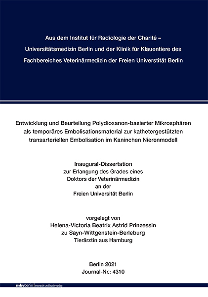
Entwicklung und Beurteilung Polydioxanon-basierter Mikrosphären als temporäres Embolisationsmaterial zur kathetergestützten transarteriellen Embolisation im Kaninchen Nierenmodell
Seiten
2021
|
1. Aufl.
Mensch & Buch (Verlag)
978-3-96729-136-0 (ISBN)
Mensch & Buch (Verlag)
978-3-96729-136-0 (ISBN)
- Keine Verlagsinformationen verfügbar
- Artikel merken
Development and evaluation of polydioxanone-based microspheres as temporary embolization material for transarterial embolization in a rabbit kidney model
The objective/aim of the present animal study was to investigate the in-vivo behavior of novel temporary embolization microspheres made of polydioxanone. It is a hypothesis-generating primary study in which the basic properties (feasibility, safety, efficacy, resorbability and biocompatibility) of the newly developed material were examined in a renal embolization model in 16 clinically healthy New Zealand White.
For this purpose, newly size-calibrated (100-150 μm and 90-315 μm) and biodegradable microspheres made of polydioxanone were developed. The selective unilateral embolization of the kidney poles was performed randomized and under fluoroscopic control. The effectiveness of the embolization was confirmed by means of digital subtraction angiography (DSA) and magnetic resonance imaging (MRI).
Three animals (group 0) were euthanized immediately after embolization in order to assess the acute behavior of the particles. The remaining 13 animals were subjected to control imaging (DSA and MRT) after 1, 4, 8, 12 or 16 weeks to assess resorbability and reperfusion. This was followed by euthanasia and laboratory processing of the target organs for the histopathological examination of the resorbability and biocompatibility of the microspheres.
Renal embolization with polydioxanone microspheres was safe to perform in all rabbits. The injection through conventional catheter systems was moderately easy. The embolization resulted in an effective vascular occlusion, which was confirmed by DSA and MRI as well as in histopathology. The DSA and MRT controls after 1, 4, 8, 12, or 16 weeks showed partial to complete reperfusion as an indication of resorbability of the microspheres. The histopathological examination confirmed the resorbability through a microscopically visible and progressive particle degradation over time. The degradation of the microspheres was accompanied by a mild to moderate inflammatory / foreign body reaction with no evidence of tissue intolerance.
In conclusion the novel temporary embolization microspheres made of polydioxanone are characterized by good applicability and safety in the rabbit kidney embolization model, as well as reliable efficacy, resorbability and biocompatibility. In order to enable clinical application, biochemical modifications to the particles with the aim of accelerated degradation behavior and improved injectability are necessary. Ziel der vorliegenden tierexperimentellen Studie war es das in-vivo Verhalten neuartiger temporärer Embolisationsmikrosphären aus Polydioxanon zu beleuchten. Es handelt sich dabei um einen hypothesen-generierenden Primärversuch, bei dem die grundlegenden Eigenschaften (Anwendbarkeit, Sicherheit, Wirksamkeit, Resorbierbarkeit und Biokompatibilität) des neu entwickelten Materials an 16 klinisch gesunden „Weißen Neuseeländern“ im Nierenembolisationsmodell untersucht wurden.
Hierzu wurden neuartige, größenkalibrierte (100 – 150 μm und 90 – 315 μm) und biodegradierbare Mikrosphären aus Polydioxanon entwickelt. Die selektive unilaterale Embolisation der Nierenpole erfolgte randomisiert und unter fluoroskopischer Kontrolle. Die Wirksamkeit der Embolisation wurde mittels digitaler Subtraktionsangiographie (DSA) und Magnetresonanztomographie (MRT) bestätigt. Drei Tiere (Gruppe 0) wurden unmittelbar nach der Embolisation euthanasiert um das Akutverhalten der Partikel zu beurteilen. Bei den übrigen 13 Tiere erfolgte nach 1, 4, 8, 12, oder 16 Wochen eine Kontrollbildgebung (DSA und MRT) zur Beurteilung der Resorbierbarkeit und Reperfusion. Anschließend erfolgte die Euthanasie und labortechnische Aufarbeitung der Zielorgane zur histopathologischen Untersuchung der Resorbierbarkeit und Biokompatibilität der Mikrosphären.
Die Nierenembolisation mit Polydioxanon-Mikrosphären lies sich in allen Kaninchen sicher durchführen. Die Injektion durch herkömmliche Kathetersysteme erfolgte dabei mäßig leicht. Die Embolisationspartikel bewerkstelligten einen wirksamen Gefäßverschluss, welcher sich sowohl mittels Angiographie und MRT, als auch in der Histopathologie bestätigte. Die Angiographie- und MRT-Kontrollen nach 1, 4, 8, 12, oder 16 Wochen zeigten eine partielle bis vollständige Reperfusion als Anzeichen einer Resorbierbarkeit der Mikrosphären. Die histopathologische Untersuchung bestätigte die Resorbierbarkeit durch eine mikroskopisch sichtbare und fortschreitende Partikeldegradation im zeitlichen Verlauf. Die Degradation der Mikrosphären wurde von einer gering- bis mittelgradigen Entzündungsreaktion/Fremdkörperreaktion ohne Anzeichen für eine Gewebeunverträglichkeit begleitet.
Bei zusammenfassender Betrachtung der vorliegenden Ergebnisse lässt sich sagen, dass neuartige temporärer Embolisationsmikrosphären aus Polydioxanon sich durch eine gute Anwendbarkeit und Sicherheit im Kaninchen Nierenembolisationsmodell, sowie eine zuverlässige Wirksamkeit, Resorbierbarkeit und Biokompatibilität auszeichnen. Um eine klinische Anwendung zu ermöglichen sind biochemische Modifikationen an den Partikeln mit dem Ziel eines beschleunigten Degradationsverhaltens und einer verbesserten Injektionsfähigkeit notwendig.
| Erscheinungsdatum | 16.12.2021 |
|---|---|
| Verlagsort | Berlin |
| Sprache | deutsch |
| Maße | 148 x 210 mm |
| Gewicht | 400 g |
| Themenwelt | Veterinärmedizin ► Allgemein |
| Veterinärmedizin ► Klinische Fächer ► Bildgebende Verfahren | |
| Veterinärmedizin ► Heimtier ► Kaninchen | |
| Veterinärmedizin ► Großtier | |
| Schlagworte | Animal Models • Arterien • Arteries • Catheterization • catheters • chirurgische Eingriffe • chirurgische Geräte • Embolie • embolism • Kaninchen • Katheter • katheterisierung • Kidneys • Materialien • Materials • microspheres (MeSH) • Mikrokugeln (MeSH) • Nieren • rabbits • surgical equipment • surgical operations • Tiermodelle • Veterinärmedizin • Veterinary Medicine |
| ISBN-10 | 3-96729-136-7 / 3967291367 |
| ISBN-13 | 978-3-96729-136-0 / 9783967291360 |
| Zustand | Neuware |
| Informationen gemäß Produktsicherheitsverordnung (GPSR) | |
| Haben Sie eine Frage zum Produkt? |
Mehr entdecken
aus dem Bereich
aus dem Bereich
Röntgenbilder sicher befunden
Buch (2025)
Thieme (Verlag)
91,00 €
Buch | Softcover (2024)
Saunders (Verlag)
166,95 €


