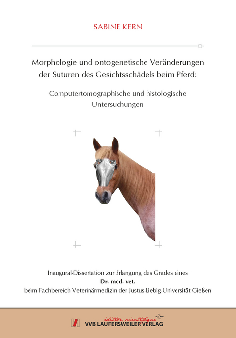
Morphologie und ontogenetische Veränderungen der Suturen des Gesichtsschädels beim Pferd: Computertomographische und histologische Untersuchungen
Seiten
2021
VVB Laufersweiler Verlag
978-3-8359-6965-0 (ISBN)
VVB Laufersweiler Verlag
978-3-8359-6965-0 (ISBN)
- Keine Verlagsinformationen verfügbar
- Artikel merken
Erkrankungen des Schädels von adulten Pferden, die mit einer primären und/oder sekundären Beteiligung von Suturen einhergehen, sind als Entzündungen der Suturen (Suturitis) und als knöcherne peri- und endostale Knochenproliferation (Suturen-Exostosen) beschrieben. Aktuelle Publikationen lassen darauf schließen, dass die Erkrankungen deutlich mehr klinische Relevanz haben als bisher angenommen. Einerseits sind die Erkrankungen lediglich bei adulten Pferden beschrieben, aber andererseits sind nur offene Suturen von diesen Erkrankungen betroffen. Diese Fakten widersprechen der bisherigen Annahme, dass die Schädelsuturen beim Pferd mit 24 Monaten knöchern durchbaut sind. Zudem fehlen morphologische Beschreibungen von Suturen des Gesichtsschädels beim Pferd, denn insbesondere diese scheinen betroffen zu sein.
Es standen insgesamt 52 CT-Datensätze von Schädeln von Pferden im Alter von unter einem Jahr bis 30 Jahren zur Verfügung. Für die Rekonstruktion der CT-Schnittbilder in 3D wurde das Programm Amira 6.2.® gewählt. Die Transversalschnittbilder aus dem CT-Datenstapel wurden von einem unerfahrenen und einem erfahrenen Untersucher unter dem Einsatz eines kommerziell erhältlichen computertomographischen Programms beurteilt und ausgewertet. Sechs Schädel wurden im Anschluss an die CT-Untersuchungen histologisch aufgearbeitet und untersucht. Beurteilt wurden die Sutura internasalis, die Sutura fronto-nasalis sinister und dexter, die Sutura lacrimo-maxillaris sinister und dexter und die Sutura zygomatico-maxillaris sinister und dexter.
Insgesamt wurden computertomographisch 135 offene, 28 partiell offene und 201 geschlossene Suturen befundet. In der histologischen Untersuchung wurden insgesamt 55 offene Suturen, die als Syndesmosen, und 346 geschlossene Suturen, die als Synostosen kategorisiert worden sind, befundet. Histomorphologisch stellten sich die Suturen folgendermaßen dar. Die Knochenränder wiesen einen zellhaltigen Saum als Übergang zu den straffen Bindegewebsfasern auf. Innerhalb des Bindegewebes waren zahlreiche, spindelförmige Fibrozyten, entweder zentral oder randständig angeordnet, und vereinzelt Blutgefäße eingelagert. Die Suturen verliefen geradlinig, mäanderförmig, herzförmig oder rund. Im Besonderen konnte ein direkter Kontakt zwischen Suturen und dem knorpeligen Septum nasi, der Schleimhaut des Sinus conchae dorsalis sowie der Nasenschleimhaut nachgewiesen werden.
Die in der Computertomographie als geschlossen befundeten Suturen wiesen auch histologisch keine Bindegewebsbrücken auf. Computertomographisch als partiell geschlossen befundete Suturen wiesen allerdings histologisch deutliche Bindegewebsbrücken zwischen den Knochenrändern auf. Histologisch wurden mehr offene Suturen vorgefunden als in der CT.
Die morphologischen Befunde wurden in Hinblick auf verschiedene Alterskategorien (jung, < 5 Jahre; adult, < 20 Jahre; geriatrisch, < 31 Jahre) analysiert. Die Sutura internasalis lag in jungen (n= 19) und adulten Pferden (n= 19) überwiegend offen (94,74%, 63,16%), in geriatrischen Pferden (n= 10) jedoch hauptsächlich partiell offen vor (80%). Sowohl die Sutura zygomatico-maxillaris als auch die Sutura lacrimo-maxillaris waren in einigen jungen Pferden (21,05%) bereits geschlossen. Nur in wenigen geriatrischen Pferden (10%) waren diese Suturen noch offen. Während sich über die Hälfte (57,89%) der Sutura fronto-nasalis in jungen Pferden bereits vollständig geschlossen hatte, stieg mit zunehmendem Alter des Pferdes die Anzahl der geschlossenen Suturen an, so dass in geriatrischen Pferden ausschließlich geschlossene Suturen vorlagen.
Die Ergebnisse dieser Studie belegen einen altersabhängigen Suturenschluss. Aus ätiopathologischer Sicht können Suturitiden oder Suturen-Exostosen nur in offenen Suturen als entzündliche Veränderung des sutureigenen Bindegewebes vorkommen.
Die vorliegende Studie dokumentiert erstmalig computertomographische und histomorphologische Charakteristika von Suturen des equinen Gesichtsschädels. Damit wird eine Basis für weiterführende, klinische Studien geschaffen. Diseases of the head in adult horses, which primarily and/or secondarily involve the sutures, are described as inflammation of the sutures (suturitis) and as peri- and endosteal bony proliferation. Current publications suggest that these diseases have more clinical relevance than previously assumed. On one hand, they are only described in adult horses but on the other hand, only open sutures are affected by these conditions.
Furthermore, morphological descriptions of the sutures in the equine facial skull are missing. These appear to be the sutures that are particularly affected. Overall, 52 CT-datasets from skulls from horses at the age of under one year to 30 years were available for investigation. The program Amira 6.2.® was used to control the sampling site and to reconstruct CT-cross-sectional images. The transverse cross-sectional images of the CT-datasets were evaluated and analyzed by an inexperienced and an experienced examiner using a commercially available computed tomography program. Following the CT examination, six skulls were processed histologically and examined. The internasal suture, the left and right fronto-nasal suture, the left and right lacrimo-maxillary suture, and the left and right zygomatico-maxillary suture were evaluated.
In total, 135 open, 28 partially open and 201 closed sutures were analyzed computer-tomographically. Overall, the result of the histological examination was a total number of 55 open sutures, classified as syndesmosis, and 346 closed sutures, categorized as synostosis. The sutures were described histomorphologically: the bone margins showed a rim of cells forming a transition to the connective tissue fibers. Numerous spindle-shaped fibrocytes, which were situated either centrally or marginally, as well as sporadic blood vessels, were embedded inside the connective tissue. The course of the sutures was linear, meander-shaped, heart-shaped or circular. Specifically, a direct contact between the sutures and the cartilaginous septum nasi, the mucosa of the sinus conchae dorsalis as well as the nasal mucosa were detected. Closed sutures in CT displayed no band of connective tissue histologically. Partially closed sutures in CT however showed a band of connective tissue between the bone margins histologically. More open sutures were found in the histological examination than by CT. Morphological results were analyzed with regard to different age categories (young, < 5 years; adult, < 20 years; geriatric, < 31 years). The internasal suture was predominantly open (94.74%, 63.16%) in young (n= 19) and adult horses (n=19), but mainly partially open (80%) in geriatric horses (n=10). The zygomatico-maxillary suture as well as the lacrimo-maxillary suture were already closed (21.05%) in some young horses. In contrast, open sutures were found only in a few geriatric horses (10%). While over half (57.89%) of the fronto-nasal suture was already completely closed in young horses, the number of closed sutures increased with increasing age of the horses, so that the sutures were already closed in geriatric horses.
The results of this study confirm an age-dependent closure of sutures. From an etiopathological standpoint, suturitis and suture exostosis can only occur in open sutures as inflammatory changes of suture-own connective tissue.
This current study describes morphological and histological characteristics of the sutures of the equine facial skull for the first time. Thus, forming a basis for further, clinical studies.
| Erscheinungsdatum | 24.06.2021 |
|---|---|
| Reihe/Serie | Edition Scientifique |
| Verlagsort | Gießen |
| Sprache | deutsch |
| Maße | 148 x 210 mm |
| Gewicht | 200 g |
| Themenwelt | Veterinärmedizin ► Allgemein |
| Veterinärmedizin ► Vorklinik ► Histologie / Embryologie | |
| Schlagworte | Doktorarbeit • Knochenbau • Tiermedizin |
| ISBN-10 | 3-8359-6965-X / 383596965X |
| ISBN-13 | 978-3-8359-6965-0 / 9783835969650 |
| Zustand | Neuware |
| Informationen gemäß Produktsicherheitsverordnung (GPSR) | |
| Haben Sie eine Frage zum Produkt? |
Mehr entdecken
aus dem Bereich
aus dem Bereich
Differentialdiagnosen beim Kleintier
Buch | Softcover (2023)
LABOKLIN GmbH & Co. KG (Verlag)
55,95 €
Grundlagen, Techniken, Präparate
Buch | Hardcover (2019)
Schlütersche (Verlag)
59,95 €


