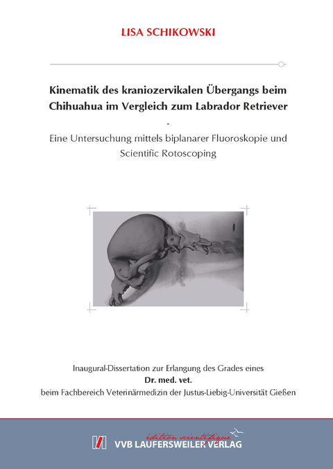
Kinematik des kraniozervikalen Übergangs beim Chihuahua im Vergleich zum Labrador Retriever.
Eine Untersuchung mittels biplanarer Fluoroskopie und Scientific Rotoscoping.
Seiten
2021
VVB Laufersweiler Verlag
978-3-8359-6921-6 (ISBN)
VVB Laufersweiler Verlag
978-3-8359-6921-6 (ISBN)
- Keine Verlagsinformationen verfügbar
- Artikel merken
Der kraniozervikale Übergang bei Zwergrassen steht auf Grund einer Reihe von Erkrankungen, die unter dem englischen Begriff „craniocervical junction abnormalities“ zusammengefasst werden (Cerda‐Gonzalez et al. 2009, Cerda-Gonzalez und Dewey 2010, Cummings et al. 2018), im Interesse der Forschung. Über die physiologischen Bewegungsabläufe des kraniozervikalen Übergangs ist bislang außer der Studie von Kelleners (2019) nichts bekannt. Kadaverstudien an herausgelösten Wirbelsäulenabschnitten oder Untersuchungen, die an anästhesierten Hunden durchgeführt wurden (Morgan et al. 1986, Penning und Badoux 1987, McLear und Saunders 2000), geben hierzu keinen Einblick. Als weiterführende Studie zu der von Kelleners (2019), in der erstmalig die Bewegungen des kraniozervikalen Übergangs beim Hund während der Lokomotion beschrieben wurden, sollen in dieser Arbeit die physiologischen Bewegungsmuster des Chihuahuas und des Labrador Retrievers während der Fortbewegung untersucht werden. Dies kann als Grundlage zur Analyse von pathologischen Beweglichkeiten hinsichtlich deren Bewegungsmustern und/oder Ausmaß in Bezug auf Erkrankungen des kraniozervikalen Übergangs genutzt werden.
Ziel der Arbeit ist eine dreidimensionale nicht invasive in vivo Bewegungsanalyse des kraniozervikalen Überganges beim klinisch gesunden Chihuahua (n=8) im Vergleich zum klinisch gesunden Labrador Retriever (n=3) in der Gangart Schritt. Der Fokus dieser Arbeit liegt sowohl auf den schrittzyklusabhängigen Bewegungen als auch auf den während der Lokomotion gezeigten aktiven Kopf/Halsbewegungen. Das Scientific Rotoscoping, als markerloses Verfahren der XROMM-Methodik (X-Ray Reconstruction of Moving Morpholoy), wurde zur kinematischen Analyse verwendet. Es setzt sich aus einer Vielzahl an einzelnen Arbeitsschritten zusammen, aus denen folgend die Bewegungsdaten entstehen. Mit Hilfe der computertomographischen Datensätze wird ein Knochenmodell erzeugt und Gelenkpunkte zwischen den einzelnen Knochen eingesetzt, sodass eine Knochenmarionette entsteht. Mit biplanarer Fluoroskopie und mit Hilfe von Highspeed-Kameras werden die Bewegungen der Hunde auf dem Laufband dokumentiert. Eine aus CT Daten generierte Knochenmarionette wird nach mehreren Bearbeitungsschritten der Röntgenvideos an diese angepasst werden. Es entsteht eine dreidimensionale, virtuelle Wirbelsäule, welche die realen Bewegungen der knöchernen Strukturen während der Fortbewegung virtuell widerspiegelt und dreidimensionale Bewegungsmessungen mit hoher Genauigkeit ermöglicht.
In der vorliegenden Arbeit konnte der bei Kelleners (2019) bereits beobachtete Zusammenhang der Rotations- und Translationsbewegungen zum Schrittzyklus bestätigt werden und zusätzlich ein Zusammenhang der lateralen Rotation der Gelenkkette, der sagittalen Rotation bei C3/C2 sowie der axialen Rotation des Atlantoaxialgelenkes beobachtet werden. Bei der biphasischen horizontalen Translation und der monophasischen lateralen Translation besteht ein Zusammenhang zu den Positionsveränderungen während der Lokomotion. Bei der vertikalen Translation, der sagittalen und axialen Rotation der Gelenkkette sowie der sagittalen Rotation von C3/C2, des Atlantookzipitalgelenkes und der axialen Rotation des Atlantoaxialgelenkes ist ein Zusammenhang zur Stemmphase und Vorschwingphase der Vordergliedmaßen erkennbar. Die schrittzyklusabhängigen Bewegungen sind jedoch nur nachvollziehbar, wenn das Muster nicht durch aktive Kopfbewegungen „gestört“ wird. Der Chihuahua und der Labrador Retriever weisen ein gleiches Grundmuster der Bewegungen auf mit individuell unterschiedlichem Amplitudenausmaß sowie geringgradig individuell variierender „Time of Occurrence“ (TOM). Es besteht eine positive Korrelation der gangzyklusabhängigen horizontalen Translation mit der vertikalen Translation sowie der vertikalen Translation mit der sagittalen Rotation der Gelenkkette. Die laterale Rotation der Gelenkkette erfolgt zeitgleich mit der lateralen Translation in gleiche Richtung. Am Ende der lateralen Translation folgt die axiale Rotation in entgegengesetzte Richtung. Der Labrador Retriever zeigt eine deutlich größere Amplitude der axialen Rotation der Gelenkkette im Vergleich zum Chihuahua.
Die gangzyklusassoziierte axiale Rotation der Gelenkkette (C3/C3) und die axiale Rotation des Atlantoaxialgelenkes zeigen eine entgegengesetzte Rotationsrichtung. Die schrittzyklusassoziierte sagittale Rotation von C3/C3 und C3/C2 weisen eine gleiche Rotationsrichtung auf mit entgegengesetzter sagittaler Rotationsrichtung des Kopfes. Der durchschnittliche Bewegungsumfang der horizontalen und vertikalen Translation sowie der axialen, lateralen und sagittalen Rotation der Gelenkkette ist beim Chihuahua kleiner als beim Labrador Retriever (horizontale Translation Ch: 0,5 ± 0,7 cm, L: 0,8 ± 0,9 cm; vertikale Translation Ch: 0,7 ± 0,9 cm, L: 0,9 ± 1,0 cm; axiale Rotation Ch: 2,6 ± 2,9°, L: 3,5 ± 3,9°; laterale Rotation Ch: 2,3 ± 2,7°, L: 2,7 ± 3,6°; sagittale Rotation Ch: 4,0 ± 5,4°, L: 4,8 ± 4,4°). Der Labrador Retriever zeigt im Vergleich zum Chihuahua eine größere Amplitude der axialen und lateralen Rotation der Gelenkkette und ein geringgradig zeitversetztes Minimum/Maximum der sagittalen Rotation nach Aktion der Vordergliedmaße. Die durchschnittliche ROM der lateralen Translation zwischen den Chihuahuas (1,0 ± 1,0 cm) und Labrador Retrievern (1,1 ± 1,9 cm) ist ungefähr gleich, ebenso wie die ROM der sagittalen Rotation bei C3/C2 (Chihuahua 2,1 ± 1,9° und Labrador Retriever 2,0 ± 1,6°). Der durchschnittliche Bewegungsumfang der axialen Rotation des Atlantoaxialgelenkes ist beim Chihuahua (4,2 ± 3,7°) geringgradig größer als beim Labrador Retriever (3,8 ± 3,6°), jedoch die ROM der sagittalen Rotation des Atlantookzipitalgelenkes des Chihuahuas (3,09 ± 3,5°) geringgradig kleiner (Labrador Retriever 3,8 ± 2,9°).
Die sagittale, laterale und axiale Rotation ist sowohl im Atlantoaxialgelenk als auch im Atlantookzipitalgelenk im Rahmen von Kopfbewegungen während der Lokomotion nachweisbar. Die laterale und axiale Rotation treten gekoppelt auf. Während der Fortbewegung werden hauptsächlich Seitwärtsbewegungen sowie Extensions- und Flexionsbewegungen des Kopfes sichtbar. Im Rahmen von aktiven Kopfbewegungen während der Lokomotion steht die sagittale Rotation im Atlantoaxial- und Atlantookzipitalgelenk mit Flexions- sowie Extensionsbewegungen im Zusammenhang. Bei einer aktiven Kopfbewegung nach links kommt es im Atlantoaxialgelenk zu einer lateralen Rotation nach links und zu einer axialen Rotation nach rechts. Im Atlantookzipitalgelenk kommt es bei dieser Kopfbewegung zu einer lateralen und axialen Rotation nach links. Im Atlantoaxialgelenk sind die größten gemessen Ranges of Motion 20° sagittale Rotation, 26° laterale Rotation und 24° axiale Rotation. Im Atlantookzipitalgelenk sind die größten gemessenen Ranges of Motion 16° laterale Rotation, 18° axiale Rotation und 30° sagittale Rotation.
Bei Betrachtung der sagittalen Rotation des Atlas zeigt der Chihuahua insgesamt durchschnittliche absolute Werte zwischen 9,1 ± 6,8° und 18,7 ± 9,9° und der Labrador Retriever zwischen 5,7 ± 4,6° und 14,5 ± 2,6°. Der Chihuahua zeigt somit im Vergleich zum Labrador Retriever durchschnittlich einen geringgradig größeren Wert der sagittalen Rotation des Atlas, welches einer steileren Achse des Atlas entspricht. Jedoch spricht die hohe Standardabweichung der Chihuahuas für eine große Varianz. Im Vergleich der ROM zwischen großen und kleinen Hunden würden bei einer Skalierung der Wirbelkörper ähnliche ROM-Werte zu erwarten sein, nicht jedoch auf Grund der unterschiedlichen Morphologie der Wirbelkörper (Angermann 2006, Schlegel et al. 2010, Spörl 2014). Da es sich in der vorliegenden Studie um zufällige Kopfbewegungen mit unterschiedlichem Bewegungsumfang zwischen den einzelnen Individuen handelt und keine standardisierten Bewegungen provoziert und gemessen werden konnten, kann nicht mit Hilfe der durchschnittlichen Werte der ROM und ROMmax zwischen den Rassen auf eine größere Beweglichkeit innerhalb einer Rotationsrichtung einer Rasse geschlossen werden. Jedoch können die absoluten Messwerte des Atlas und des Schädels im Vergleich zur Referenzposition, der CT-Lagerung, einen Hinweis auf unterschiedliche Winkelungen der Wirbelkörper zwischen beiden Rassen während der Lokomotion geben.
Im Interobserver-Vergleich mit Kelleners (2019) zeigt sich eine hohe Übereinstimmung der erhobenen Daten bei Betrachtung der Translationsbewegungen der Gelenkkette. Ein gleiches Grundmuster der schrittzyklusabhängigen Rotationsbewegungen und aktiven Kopfbewegungen ist ebenfalls zwischen beiden Anwendern erkennbar, jedoch variieren hier Ausmaß, Dauer und Zeitpunkt vor allem von kleineren Bewegungen stärker.
Die Ergebnisse dieser Studie bestätigen den Zusammenhang von Kopf- und Halsbewegungen mit der Fortbewegung und zeigen erstmals ein gleiches Muster dieser Bewegungen bei Labrador Retrievern und Chihuahuas. Die zyklisch auftretenden Positionsveränderungen und die Aktion der Vordergliedmaßen verursachen die Kopf- und Halsbewegungen. Diese können von aktiven Bewegungen im kraniozervikalen Übergang überlagert werden. Die axiale Rotationsbewegung und deren Kopplung mit der lateralen Rotation im Atlantookzipitalgelenk, die Kelleners (2019) erstmalig beschrieben hat, konnte ebenfalls bestätigt werden. Zusätzlich wird erstmals die schrittzyklusabhängige laterale Rotation der Gelenkkette sowie die schrittzyklusabhängige axiale Rotation des Atlantoaxialgelenkes beschrieben. Des Weiteren wurden physiologische Messwerte der Range of Motion während der Lokomotion für die sagittale, laterale und axiale Rotation bei beiden untersuchten Rassen bestimmt.
Die vorliegende Arbeit liefert damit Erkenntnisse zur dreidimensionalen Bewegung des kraniozervikalen Übergangs während der Lokomotion beim Chihuahua und Labrador Retriever und der dort spontan auftretenden aktiven Kopfbewegungen. Sie schafft eine Grundlage für weitere Vergleichsstudien und liefert einen Beitrag zum besseren Verständnis der physiologischen Gegebenheiten und Variationen zwischen den beiden untersuchten Rassen. The craniocervical junction in toy breeds is in the interest of research due to a variety of diseases that are summarized under the term "craniocervical junction abnormalities" (Cerda‐Gonzalez et al. 2009, Cerda-Gonzalez und Dewey 2010, Cummings et al. 2018). Apart from the study by Kelleners (2019), nothing is known so far about the physiological movement patterns of the craniocervical junction. Cadaver studies on detached spinal column sections or studies performed on anaesthetized dogs (Morgan et al. 1986, Penning und Badoux 1987, McLear und Saunders 2000) do not provide any insight into this topic. Kelleners (2019) first described the movement patterns of the craniocervical junction in dogs during locomotion, while in this study, as a further research to Kelleners (2019), the physiological movement patterns of the Chihuahua and Labrador Retriever during locomotion will be examined. This can serve as a basis for the analysis of pathological movement patterns and/or their extent in relation to diseases of the craniocervical junction.
The aim of this study is a three-dimensional non-invasive in vivo motion analysis of the craniocervical junction of clinically sound Chihuahuas (n=8) compared to clinically sound Labrador Retrievers (n=3). The focus of this study is on gait-cycle-related movements while walking as well as active head/neck movements during locomotion. “Scientific Rotoscoping”, a markerless method of the XROMM methodology (X-Ray Reconstruction of Moving Morphology), was used for kinematic analysis. It is composed of a large number of individual work steps, from which the movement data are subsequently generated. For this purpose, a bony marionette is constructed based on computer tomographic data sets and articular joints are inserted. Biplanar fluoroscopy and high-speed cameras are used to document the dog’s movements on the treadmill. After several processing steps the bone marionette is matched with the bony silhouette on X-ray videos according to the movement observed. A three-dimensional, virtual spinal column is then created which virtually reflects the real movement patterns of the bony structures during locomotion and enables three-dimensional movement measurements with high accuracy.
In the present study the relationship between the rotational and translational movements and the gait cycle, already observed in Kelleners (2019), was confirmed. Additionally, a relationship between the lateral rotation of the bone marionette, the sagittal rotation at C3/C2 and the axial rotation of the atlantoaxial joint was also determined. There is a correlation between the biphasic horizontal translation and the monophasic lateral translation and the changes in position during locomotion. The vertical translation, the sagittal and axial rotation of the bone marionette as well as the sagittal rotation of C3/C2, the atlantooccipital joint and the axial rotation of the atlantoaxial joint show a relation to the stem phase and swing phase of the forelimbs. However, the gait cycle-dependent movements are only visible if the pattern is not "disturbed" by active head movements. The Chihuahua and the Labrador Retriever have the same basic pattern of movements with individually varying amplitude and slightly varying "Time of Occurrence" (TOM). There is a positive correlation between the gait cycle-dependent horizontal translation and the vertical translation as well as between the vertical translation and the sagittal rotation of the bone marionette. The lateral rotation of the bone marionette occurs simultaneously with the lateral translation in the same direction. At the end of the lateral translation the axial rotation occurs in opposite direction. The Labrador Retriever shows a significantly greater amplitude of axial rotation of the bone marionette compared to Chihuahuas. The gait-cycle-associated axial rotation of the bone marionette (C3/C3) and the axial rotation of the atlantoaxial joint show an opposite direction of rotation. The gait-cycle-associated sagittal rotation of C3/C3 and C3/C2 show the same direction of rotation with opposite sagittal rotation of the head. The average range of motion of the horizontal and vertical translation and the axial, lateral and sagittal rotation of the bone marionette is smaller in Chihuahuas than in Labrador Retrievers (horizontal translation Ch: 0.5 ± 0.7 cm, L: 0.8 ± 0.9 cm; vertical translation Ch: 0.7 ± 0.9 cm, L: 0.9 ± 1.0 cm; axial rotation Ch: 2.6 ± 2.9°, L: 3.5 ± 3.9°; lateral rotation Ch: 2.3 ± 2.7°, L: 2.7 ± 3.6°; sagittal rotation Ch: 4.0 ± 5.4°, L: 4.8 ± 4.4°). Compared to Chihuahuas, Labrador Retrievers show a greater amplitude of axial and lateral rotation of the bone marionette and a slightly time-delayed minimum/maximum of sagittal rotation after action of the forelimb. The average ROM of lateral translation between Chihuahuas (1.0 ± 1.0 cm) and Labrador Retrievers (1.1 ± 1.9 cm) is approximately the same, as is the ROM of sagittal rotation at C3/C2 (Chihuahua 2.1 ± 1.9° and Labrador Retriever 2.0 ± 1.6°). The average range of motion of axial rotation of the atlantoaxial joint is slightly greater in Chihuahuas (4.2 ± 3.7°) than in Labrador Retriever (3.8 ± 3.6°), but the ROM of sagittal rotation of the atlantooccipital joint of Chihuahuas (3.09 ± 3.5°) is slightly smaller (Labrador Retriever 3.8 ± 2.9°).
When moving, the sagittal, lateral and axial rotation can be detected in both, the atlantoaxial and the atlantooccipital joint during head movements. Lateral and axial rotation occur as a coupled motion pattern. In locomotion mainly lateral movements as well as extension and flexion movements of the head become visible. During active head movements in locomotion, sagittal rotation in the atlantoaxial and atlantooccipital joint is related to flexion and extension movements. When moving the head actively to the left, lateral rotation to the left and axial rotation to the right occurs in the atlantoaxial joint. In the atlantooccipital joint this head movement results in a lateral and axial rotation to the left. In the atlantoaxial joint the widest measured ranges of motion are 20° sagittal rotation, 26° lateral rotation and 24° axial rotation. In the atlantooccipital joint, the widest measured ranges of motion are 16° lateral rotation, 18° axial rotation and 30° sagittal rotation.
Looking at the sagittal rotation of the atlas, Chihuahuas show, on the whole, average absolute values between 9.1 ± 6.8° and 18.7 ± 9.9° and the Labrador Retriever between 5.7 ± 4.6° and 14.5 ± 2.6°. Thus, Chihuahuas show a slightly higher average value of the sagittal rotation of the atlas compared to Labrador Retrievers, which corresponds to a steeper axis of the atlas. However, the high standard deviation of the Chihuahua indicates a large variance. Comparing the ROM between large and small dogs, similar ROM values would be expected if the vertebral bodies were scaled, but not due to the different morphology of the vertebral bodies (Angermann 2006, Schlegel et al. 2010, Spörl 2014). The present study involves random head movements with varying degrees of movement between individuals and no standardized movements could be provoked and measured. Therefore, the average values of ROM and ROMmax between breeds cannot be used to assume a greater mobility within a direction of rotation of a breed. However, in locomotion the absolute measured values of the atlas and the skull in comparison to the reference position, the CT position, can give an indication of different angulations of the vertebral bodies between both breeds of dogs.
The interobserver comparison shows that the collected data coincides with the data from Kelleners (2019) when considering the translational movements of the bone marionette. An identical basic movement pattern of step-cycle-dependent rotational movements and active head movements can also be seen between the two users, but the extent, duration and timing especially of small movements vary to a greater degree.
The results of this study confirm the correlation between head and neck movements and locomotion. For the first time a similar pattern of these movements is shown in the Labrador Retriever and Chihuahua. General cyclical position changes at walk as well as movements of the forelimbs have an effect on the head-neck movement in locomotion, when the craniocervical junction is not actively moved. The axial rotational movement and its coupling with the lateral rotation in the atlantooccipital joint, which was first described by Kelleners (2019), could also be confirmed. In addition, the step-cycle-dependent lateral rotation of the bone marionette and the step-cycle-dependent axial rotation of the atlantoaxial joint are described for the first time. Furthermore, physiological measured values of the range of motion during locomotion were determined for the sagittal, lateral and axial rotation in both breeds of dogs.
The present study gives insights into the three-dimensional kinematics of the craniocervical joints in locomotion and in active head movements of Chihuahuas and Labrador Retrievers. It provides a basis for further comparative studies and contributes to a better understanding of the physiological conditions and variations between the two breeds of dogs studied.
| Erscheinungsdatum | 01.03.2021 |
|---|---|
| Reihe/Serie | Edition Scientifique |
| Sprache | deutsch |
| Maße | 148 x 210 mm |
| Gewicht | 860 g |
| Themenwelt | Veterinärmedizin ► Allgemein |
| Veterinärmedizin ► Vorklinik | |
| Veterinärmedizin ► Kleintier | |
| Schlagworte | Hund • Promotion • Tiermedizin |
| ISBN-10 | 3-8359-6921-8 / 3835969218 |
| ISBN-13 | 978-3-8359-6921-6 / 9783835969216 |
| Zustand | Neuware |
| Haben Sie eine Frage zum Produkt? |

