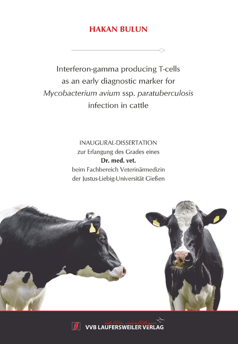
Interferon-gamma producing T-cells as an early diagnostic marker for Mycobacterium avium ssp. paratuberculosis infection in cattle
Seiten
2020
VVB Laufersweiler Verlag
978-3-8359-6901-8 (ISBN)
VVB Laufersweiler Verlag
978-3-8359-6901-8 (ISBN)
- Keine Verlagsinformationen verfügbar
- Artikel merken
Bovine paratuberculosis is a chronic intestinal infection caused by Mycobacterium avium ssp. paratuberculosis (MAP). Early detection of subclinical paratuberculosis is crucial as calves, exposed to MAP early in life, are highly susceptible to acquire the infection. In early stage of infection, infected animals develop an effective cell-mediated immune response to MAP-antigens with an increase in interferon-gamma (IFN-γ) production. Quantification of extracellular IFN-γ in antigen-stimulated whole blood samples often results in false positive reactions when applied to juvenile animals. The present study aimed to improve the performance of a method suggested by Waters et al. (2003a) deploying flow cytometry analysis (FCA) to quantify antigen-induced IFN-γ production at the level of single cells. To this end, different antigen preparations for in vitro-restimulation and different methods of data analysis were assessed. Six MAP-negative calves and 6 calves orally inoculated with MAP at 10 days of age were sampled every 4 weeks for 52 weeks post inoculation (w.p.i.). Peripheral blood mononuclear cells (PBMC) were stimulated with either purified protein derivatives (PPD) or whole cell sonicates (WCS) derived from MAP, M. avium ssp. avium (MAA) or M. phlei (MP), respectively, for 6 days followed by labeling of intracellular IFN-γ in CD4+ and in CD8+ T-cells. By FCA, lymphocytes and lymphoblasts were separately analysed. No antigen-specific IFN-γ production was detectable in CD8+ T-cells throughout and the responses of CD4+ lymphocytes and lymphoblasts of MAP-infected and control calves were similar up to 12 w.p.i. Later on, however, the mean fluorescence intensity (MFI) for the detection of IFN-γ in CD4+ lymphocytes and lymphoblasts after WCS-MAP antigen stimulation allowed for a differentiation of animal groups. Difference became significant as early as 16 w.p.i. and remained significantly different until the end of the sampling period (except at 44 w.p.i.). CD4+ T-cells of 4/6 MAP-infected calves significantly responded to WCS-MAP 16 w.p.i. During the whole observation period, at least three samples of any of the infected calves tested positive. This diagnostic approach was calculated to have a superior sensitivity (87.8%) and specificity (86.8%) to detect infected animals in an early stage of the MAP infection as compared to the IFN-γ release assay. The quantification of MAP-antigen specific IFN-γ production at the level of individual CD4+ T-cells via FCA may serve as a valuable diagnostic tool to identify MAP-infected juvenile cattle.
| Erscheinungsdatum | 17.11.2020 |
|---|---|
| Reihe/Serie | Edition Scientifique |
| Sprache | englisch |
| Maße | 146 x 210 mm |
| Gewicht | 200 g |
| Themenwelt | Veterinärmedizin |
| Schlagworte | Doktorarbeit • Uni • Wissenschaft |
| ISBN-10 | 3-8359-6901-3 / 3835969013 |
| ISBN-13 | 978-3-8359-6901-8 / 9783835969018 |
| Zustand | Neuware |
| Informationen gemäß Produktsicherheitsverordnung (GPSR) | |
| Haben Sie eine Frage zum Produkt? |
Mehr entdecken
aus dem Bereich
aus dem Bereich
A Practical Guide
Buch | Hardcover (2024)
Wiley-Blackwell (Verlag)
145,95 €
Buch | Hardcover (2024)
Wiley-Blackwell (Verlag)
124,55 €
Buch | Softcover (2024)
British Small Animal Veterinary Association (Verlag)
87,25 €


