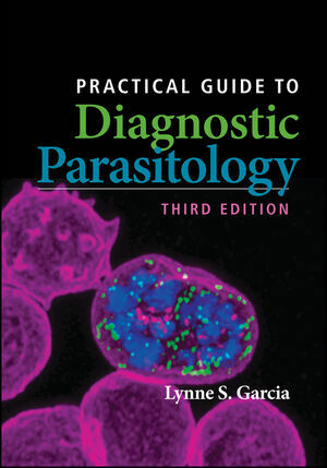
Practical Guide to Diagnostic Parasitology
American Society for Microbiology (Verlag)
978-1-68367-039-1 (ISBN)
Specimen collection, preservation, and testing options are thoroughly discussed, from the routine ova and parasite examination to blood films, fecal immunoassays, and the newer molecular test panels. Specific test procedures, laboratory methods and reagents, and algorithms are provided. The ever-helpful "FAQ" section of commonly asked questions now offers expanded information on stool specimen fixatives and testing, thorough coverage of new techniques, and advice on reporting and commenting on results.
The heart of the Guide, covering identification of individual pathogens, has been expanded with more discussion and comparison of organisms and dozens of new color images. An entirely new section has been added that uses extensive figures and new tables to illustrate common problems with differentiating organisms from one another and from possible microscopic artifacts. The final section has been reorganized to include identification keys and dozens of tables summarizing organism characteristics to assist the bench microbiologist with routine diagnostic testing methods.
Lynne Shore Garcia is the director of LSG & Associates, a firm providing training, teaching, and consultation services for diagnostic medical parasitology and health care administration. A former manager of the UCLA Clinical Microbiology Laboratory, she is a sought-after speaker (nationally and internationally) and author of hundreds of articles, book chapters, and books including two ASM Press books, Clinical Laboratory Management, Second Edition and Diagnostic Medical Parasitology, Sixth Edition.
Garcia 3e TOC draft from mss
Preface 000
Acknowledgments 000
SECTION 1 Philosophy and Approach to Diagnostic Parasitology
Neglected Tropical Diseases
Why Perform Diagnostic Parasitology Testing?
Travel
Population Movements
Control Issues
Global Climate Change
Epidemiologic Considerations
Compromised Patients; Potential Sex Bias Regarding Infection Susceptibility, Aging
Approach to Therapy
Who Should Perform Diagnostic Parasitology Testing?
Laboratory Personnel
Nonlaboratory Personnel
Where Should Diagnostic Parasitology Testing Be Performed?
Inpatient Setting
Outpatient or Referral Setting
Decentralized Testing
Physician Office Laboratories
Over-the-Counter (Home Care) Testing
Field Sites
What Factors Should Precipitate Testing?
Travel and Residence History
Immune Status of the Patient
Clinical Symptoms
Documented Previous Infection
Contact with Infected Individuals
Potential Outbreak Testing
Occupational Testing
Therapeutic Failure
What Testing Should Be Performed?
Routine Tests
Special Testing, Reference Laboratories
Specialized Referral Test Options: DPDx and Other Sites
Other (Nonmicrobiological) Testing
What Factors Should Be Considered When Developing Test Menus?
Physical Plant
Client Base
Customer Requirements and Perceived Levels of Service
Personnel Availability and Level of Expertise
Equipment
Budget
Risk Management Issues Associated with Stat Testing
Primary Amebic Meningoencephalitis (PAM)
Granulomatous Amebic Encephalitis and Amebic Keratitis
Request for Blood Films
Automated Instrumentation
Patient Information
Conventional Microscopy
SECTION 2 Parasite Classification and Relevant Body Sites
Protozoa (Intestinal)
Amebae, Stramenopiles
Flagellates
Ciliates
Apicomplexa (Including Coccodia)
Microsporidia (Now Classified with the Fungi)
Protozoa (Other Body Sites)
Amebae
Flagellates
Apicomplexa (Including Coccidia)
Microsporidia (Now Classified with the Fungi)
Protozoa (Blood and Tissue)
Apicomplexa (Including Sporozoa)
Flagellates
Leishmaniae
Trypanosomes
Nematodes (Intestinal)
Nematodes (Tissue)
Nematodes (Blood and Tissue)
Cestodes (Intestinal)
Cestodes (Tissue)
Trematodes (Intestinal)
Trematodes (Liver and Lungs)
Trematodes (Blood)
Pentastomids
Acanthocephala
Table 2.1 Classification of human parasites
Table 2.2 Cosmopolitan distribution of common parasitic infections (North America, Mexico, Central America, South America, Europe, Africa, Asia, and Oceania)
Table 2.3 Body sites and possible parasites recovered (trophozoites, cysts, oocysts, spores, adults, larvae, eggs, amastigotes, and trypomastigotes)
SECTION 3 Collection Options
Safety
Collection of Fresh Stool Specimens
Collection Method
Number of Specimens To Be Collected
Standard Approach
Different Approaches
Collection Times
Posttherapy Collection
Specimen Type, Stability, and Need for Preservation
Preservation of Stool Specimens
Overview of Preservatives
Formalin
Sodium Acetate-Acetic Acid-Formalin (SAF)
Schaudinn's Fluid
Schaudinn's Fluid containing PVA (mercury base)
Schaudinn's Fluid containing PVA (copper base, zinc base)
Single-Vial Collection Systems (Other Than SAF)
Universal Fixative (TOTAL-FIX)
Quality Control for Preservatives
Procedure Notes for Use of Preservatives (Stool Fixative Collection Vials)
Procedure Limitations for Use of Preservatives (Stool Fixative Collection Vials)
Collection of Blood
Collection and Processing
Stat Test Requests and Risk Management Issues
Collection of Specimens from Other Body Sites
Table 3.1 Fecal specimens for parasites: options for collection and processinga
Table 3.2 Approaches to stool parasitology: test ordering
Table 3.3 Preservatives and procedures commonly used in diagnostic parasitology (stool specimens)
Table 3.4 Advantages of thin and thick blood films
Table 3.5 Advantages and disadvantages of buffy coat films
Table 3.6 Potential problems of using EDTA anticoagulant for the preparation of thin and thick blood films
Table 3.7 Body sites and possible parasites recovered (trophozoites, cysts, oocysts, spores, adults, larvae, eggs, amastigotes, and trypomastigotes)
SECTION 4 Specimen Test Options: Routine Diagnostic Methods and Body Sites
Ova and Parasite Examination of Stool Specimens
Other Diagnostic Methods for Stool Specimens
Culture of Larval-Stage Nematodes
Estimation of Worm Burdens through Egg Counts
Hatching Test for Schistosome Eggs
Screening Stool Samples for Recovery of a Tapeworm Scolex
Testing of Other Intestinal Tract Specimens
Examination for Pinworm
Sigmoidoscopy Material
Duodenal Drainage Material
Duodenal Capsule Technique (Entero-Test)
Urogenital Tract Specimens
Sputum
Aspirates
Biopsy Specimens
Blood
Thin Blood Films
Thick Blood Films
Blood Staining Methods
Buffy Coat Films
QBC Microhematocrit Centrifugation Method
Knott Concentration
Membrane Filtration Technique
Culture Methods
Animal Inoculation and Xenodiagnosis
Antibody and Antigen Detection
Antibody Detection
Antigen Detection, Nucleic Acid-Based Tests, and Molecular Panels
Intradermal Tests
Table 4.1 Body site, procedures and specimens, recommended methods and relevant parasites, and comments
Table 4.2 Serologic, antigen, and probe tests used in the diagnosis of parasitic infections
SECTION 5 Specific Test Procedures and Algorithms
Microscopy
CALIBRATION OF THE MICROSCOPE
Ova and Parasite Examination
DIRECT WET FECAL SMEAR
Concentration (Sedimentation and Flotation)
SEDIMENTATION CONCENTRATION (Formalin-Ethyl Acetate)
SEDIMENTATION CONCENTRATION USING THE UNIVERSAL FIXATIVE (TOTAL-FIX)
FLOTATION CONCENTRATION (Zinc Sulfate)
Permanent Stained Smear
PREPARATION OF MATERIAL FOR STAINING
Fresh Material
Preserved Material Containing PVA
SAF-Preserved Material
Universal Fixative (Total-Fix) Preserved Material
Alternative Method for Smear Preparation Directly from Vial
(Total-Fix)
Stains Used in the Permanent Stained Smear
TRICHROME STAIN (Wheatley's Method)
IRON HEMATOXYLIN STAIN (Spencer-Monroe Method)
IRON HEMATOXYLIN STAIN (Tompkins-Miller Method)
MODIFIED IRON HEMATOXYLIN STAIN (Incorporating the Carbol Fuchsin Step)
POLYCHROME IV STAIN
CHLORAZOL BLACK E STAIN
Specialized Stains for Coccidia and Microsporidia
KINYOUN'S ACID-FAST STAIN (Cold Method)
MODIFIED ZIEHL-NEELSEN ACID-FAST STAIN (Hot Method)
CARBOL FUCHSIN NEGATIVE STAIN FOR CRYPTOSPORIDIUM (W. L. Current)
RAPID SAFRANIN METHOD FOR CRYPTOSPORIDIUM (D. Baxby)
RAPID SAFRANIN METHOD FOR CYCLOSPORA, USING A MICROWAVE OVEN (Govinda Visvesvara)
AURAMINE O STAIN FOR COCCIDIA (Thomas Hanscheid)
MODIFIED TRICHROME STAIN FOR MICROSPORIDIA (Weber, Green Counterstain)
MODIFIED TRICHROME STAIN FOR MICROSPORIDIA (Ryan, Blue Counterstain)
MODIFIED TRICHROME STAIN FOR MICROSPORIDIA (Evelyn Kokoskin, Hot Method)
Fecal Immunoassays for Intestinal Protozoa
Entamoeba histolytica
Cryptosporidium spp.
Giardia lamblia
Kits under Development
Comments on the Performance of Fecal Immunoassays
Enzyme Immunoassays (Antigen Detection, No Centrifugation Recommended)
Fluorescence (Visual Identification of the Organisms, Centrifugation Recommended)
Lateral-Flow Cartridges (Antigen Detection, No Centrifugation Recommended)
Larval Nematode Culture
HARADA-MORI FILTER PAPER STRIP CULTURE
BAERMANN CONCENTRATION
AGAR PLATE CULTURE FOR STRONGYLOIDES STERCORALIS
Other Methods for Gastrointestinal Tract Specimens
EXAMINATION FOR PINWORM (Cellulose Tape Preparations)
SIGMOIDOSCOPY SPECIMENS (Direct Wet Smear)
SIGMOIDOSCOPY SPECIMENS (Permanent Stained Smear)
DUODENAL ASPIRATES
Methods for Urogenital Tract Specimens
RECEIPT OF DRY SMEARS
DIRECT SALINE MOUNT
PERMANENT STAINED SMEAR
URINE CONCENTRATION (Centrifugation)
URINE CONCENTRATION (Nuclepore Membrane Filter)
Preparation of Blood Films
THIN BLOOD FILMS
THICK BLOOD FILMS
COMBINATION THICK-THIN BLOOD FILMS
RISK MANAGEMENT ISSUES ASSOCIATED WITH BLOOD FILMS
USE OF A REFERENCE LABORATORY FOR PARASITE BLOOD DIAGNOSTIC TESTING
BLOOD FILM REPORTING WITH ADDITIONAL REPORT COMMENTS
BUFFY COAT BLOOD FILMS
Blood Stains
STAIN OPTIONS
GIEMSA STAIN
Blood Concentration
BUFFY COAT CONCENTRATION
KNOTT CONCENTRATION
MEMBRANE FILTRATION CONCENTRATION
Algorithm 5.1 Procedure for processing fresh stool for the O&P examination
Algorithm 5.2 Procedure for processing liquid specimens for the O&P examination
Algorithm 5.3 Procedure for processing preserved stool for the O&P examination-two-vial collection kit
Algorithm 5.4 Procedure for processing SAF-preserved stool for the O&P examination
Algorithm 5.5 Procedure for the use of Total-Fix (Universal Fixative, single vial system) (Alternate Method for Smear Preparation Directly from Vial)
Algorithm 5.6 Use of various fixatives and their recommended stains
Algorithm 5.7 Ordering algorithm for laboratory examination for intestinal parasites
Algorithm 5.8 Procedure for processing blood specimens for examination
Table 5.1 Body site, specimen, and recommended stain(s)
Table 5.2 Approaches to stool parasitology: test ordering
Table 5.3 Laboratory test reports: optional comments
Table 5.4 Parasitemia determined from conventional light microscopy: clinical correlation
Section 6 Commonly Asked Questions about Diagnostic Parasitology
Stool Parasitology
Specimen Collection
Intestinal Tract
Fixatives
Specimen Processing
O&P Exam
Diagnostic Methods
Direct Wet Examinations
Concentrations
Permanent Stains
Stool Immunoassay Options
Molecular Test Panels (FDA Approved)
APTIMA Trichomonas vaginalis Assay
Affirm VPIII Microbial Identification Test
Cepheid Xpert TV Assay for Trichomonas vaginalis from men and women
BD MAX Enteric Parasite Panel
BioFire FilmArray Gastrointestinal panel
Luminex (VERIGENE II GI Flex Assay, includes Parasites)
Other Pending Molecular Tests
Organism Identification
Protozoa
Helminths
Reporting
Organism Identification
Quantitation
Proficiency Testing
Wet Preparations
Permanent Stained Smears
Tissues or Fluids
Blood
Specimen Collection
Specimen Processing
Diagnostic Methods
Organism Identification
Reporting
Proficiency Testing
General Questions
SECTION 7 Parasite Identification
Protozoa
Amebae (Intestinal)
Entamoeba histolytica
Entamoeba histolytica/Entamoeba dispar
Comments on Entamoeba moshkovskii and Entamoeba bangladeshi
Entamoeba bangladeshi
Entamoeba hartmanni
Entamoeba coli
Entamoeba gingivalis
Entamoeba polecki
Endolimax nana
Iodamoeba butschlii
Blastocystis spp. (formerly Blastocystis hominis)
Flagellates (Intestinal)
Giardia lamblia (G. duodenalis, G. intestinalis)
Dientamoeba fragilis
Chilomastix mesnili
Pentatrichomonas hominis
Enteromonas hominis, Retortamonas intestinalis
Ciliates (Intestinal)
Balantidium coli
Apicomplexa (Intestinal)
Cryptosporidium spp.
Coccidia (Intestinal)
Cyclospora cayetanensis
Cystoisospora (Isospora) belli
Microsporidia (Intestinal)
Enterocytozoon bieneusi
Encephalitozoon intestinalis, Encephalitozoon spp.
Sporozoa (Blood and Tissue)
Plasmodium vivax
Plasmodium falciparum
Plasmodium malariae
Plasmodium ovale wallickeri, Plasmodium ovale curtisi
Plasmodium knowlesi
Malaria: Key Diagnostic Points
Babesia spp. (B. microti, B. duncani, B. divergens, B. venatorum)
Toxoplasma gondii
Flagellates (Blood and Tissue)
Leishmania spp.
Trypanosoma brucei gambiense (West), T. brucei rhodesiense (East)
Trypanosoma cruzi
Amebae (Other Body Sites)
Naegleria fowleri
Acanthamoeba spp., Balamuthia mandrillaris, Sappinia diploidea
Flagellates (Other Body Sites)
Trichomonas vaginalis
NEMATODES
Intestinal
Ascaris lumbricoides
Trichuris trichiura;General Comments on Capillaria philippinensis
Necator americanus, Ancylostoma duodenale, A. ceylanicum (Hookworms); General comments on Trichostrongylus spp.
Strongyloides stercoralis
Enterobius vermicularis
Tissue
Ancylostoma braziliense, Ancylostoma caninum, Uncinaria stenocephala (Dog and Cat Hookworms)
Toxocara canis, Toxocara cati (Dog and Cat Ascarid Worms)
Dracunculus medinensis
Trichinella spiralis
Blood and Tissue
Filarial Worms
CESTODES
Intestinal
Taenia saginata
Taenia solium
Diphyllobothrium latum
Hymenolepis (Rodentolepis) nana
Hymenolepis diminuta
Dipylidium caninum
Tissue
Echinococcus granulosus, E. multilocularis, E. vogeli, E. oligarthrus
TREMATODES
Intestinal
Fasciolopsis buski
Liver and Lungs
Paragonimus westermani, Paragonimus mexicanus, Paragonimus kellicotti
Fasciola hepatica
Clonorchis sinensis (Chinese liver fluke) (Opisthorchis sinensis)
Blood
Schistosoma spp. (S. mansoni, S. haematobium, S. japonicum, S. mekongi, S. malayensis, S. intercalatum)
SECTION 8 Common Problems in Parasite Identification
Table 8.1 Entamoeba spp., trophozoites versus macrophages
Table 8.2 Entamoeba spp., cysts versus polymorphonuclear leukocytes
Table 8.3 Entamoeba histolytica versus Entamoeba coli precysts and cysts
Table 8.4 Endolimax nana versus Dientamoeba fragilis
SECTION 9 Identification Aids
Diagnostic Considerations
Table 9.1 Rapid diagnostic procedures
Table 9.2 Diagnostic characteristics for organisms in wet mounts (direct or concentration sediment)
Table 9.3 Diagnostic characteristics for organisms in permanent stained smears
Key 9.1 Identification of intestinal amebae (permanent stained smear)
Key 9.2 Identification of intestinal flagellates
Key 9.3 Identification of helminth eggs
Key 9.4 Identification of microfilariae
Protozoa
Table 9.4 Intestinal protozoa: trophozoites of common amebae
Table 9.5 Intestinal protozoa: cysts of common amebae
Table 9.6 Intestinal protozoa: trophozoites of less common amebae
Table 9.7 Intestinal protozoa: cysts of less common amebae
Table 9.8 Morphologic criteria used to identifyBlastocystisspp.
Table 9.9 Intestinal protozoa: trophozoites of flagellates
Table 9.10 Intestinal protozoa: cysts of flagellates
Table 9.11 Intestinal protozoa: ciliates
Table 9.12 Apicomplexa
Table 9.13 Microsporidia (related to the fungi), general information
Table 9.14 Microsporidia: recommended diagnostic techniques
Table 9.15 Comparison of Naegleria fowleri, Acanthamoeba spp., Balamuthia mandrillaris, and Sappinia diploidea
Table 9.16 Characteristics of Trichomonas vaginalis
Table 9.17 Key characteristics of intestinal tract/urogenital system protozoa
Helminths
Table 9.18 Normal life spans of the most common intestinal nematodes
Table 9.19 Characteristics of the most common intestinal nematodes
Table 9.20 Tissue nematodes
Table 9.21 Trichinella spiralis: life cycle stages and clinical conditions
Table 9.22 Characteristics of human microfilariae
Table 9.23 Characteristics of cestode parasites (intestinal)
Table 9.24 Tissue cestodes
Table 9.25 Characteristics of intestinal trematodes
Table 9.26 Characteristics of liver and lung trematodes
Table 9.27 Human paragonimiasis
Table 9.28 Characteristics of blood trematodes
Table 9.29 Key characteristics of helminths
Blood Parasites
Table 9.30 Malaria characteristics with fresh blood or blood collected using EDTA with no extended lag time
Table 9.31 Potential problems using EDTA anticoagulant for the preparation of thin and thick blood films
Table 9.32 Plasmodia in Giemsa-stained thin blood smears
Table 9.33 Relevant issues for handling requests for blood parasite infections
Table 9.34 Features of human leishmanial infectionsa
Table 9.35 Characteristics of American trypanosomiasis
Table 9.36 Characteristics of East and West African trypanosomiasis
Table 9.37 Key characteristics of blood parasites
Index
| Erscheinungsdatum | 07.06.2021 |
|---|---|
| Verlagsort | Washington DC |
| Sprache | englisch |
| Maße | 181 x 252 mm |
| Gewicht | 1236 g |
| Einbandart | kartoniert |
| Themenwelt | Medizin / Pharmazie ► Medizinische Fachgebiete |
| Naturwissenschaften ► Biologie | |
| Veterinärmedizin | |
| ISBN-10 | 1-68367-039-6 / 1683670396 |
| ISBN-13 | 978-1-68367-039-1 / 9781683670391 |
| Zustand | Neuware |
| Informationen gemäß Produktsicherheitsverordnung (GPSR) | |
| Haben Sie eine Frage zum Produkt? |
aus dem Bereich


