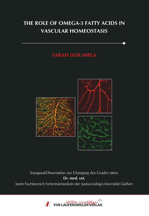
The role of omega-3 fatty acids in vascular homeostasis
Seiten
2018
VVB Laufersweiler Verlag
978-3-8359-6660-4 (ISBN)
VVB Laufersweiler Verlag
978-3-8359-6660-4 (ISBN)
- Keine Verlagsinformationen verfügbar
- Artikel merken
Polyunsaturated fatty acids (PUFAs) are organic acids, which possess more than one double bond in their carbon long chains. Biologically relevant families are the ω-3 and ω-6 fatty acids which can be metabolized by different pathways including the cyclooxygenases and lipoxygenases. Besides these oxygenases also the Cytochrome P450 (CYP) enzymes metabolize PUFAs to generate PUFA epoxides – and are frequently referred to as the “third” PUFA metabolic pathway. The most studied epoxides are the ω-6 derived epoxyeicosatrieonic acids (EETs), which demonstrate angiogenic, cardioprotective and anti-inflammatory properties. The bioactive epoxides formed are further metabolized by the soluble epoxide hydrolase (sEH) to their corresponding diols – which may have either similar or distinctly different biological activity than the epoxides.
Recently, our group discovered that the ω-3 fatty acid derived lipid mediator 19,20-dihydroxydocosapentaenoic acid (19,20-DHDP) is required for postnatal retinal angiogenesis. As it is important for retinal angiogenesis one aim of this study was to investigate the effect of the ω-3 PUFA 19,20-DHDP on angiogenesis and lymphangiogenesis compared to other PUFA metabolites. We analyzed the formation of lymphatic endothelial cells (LECs) and blood endothelial cells (BECs) in in vivo experiments (Matrigel-plug assay) and in ex vivo experiments by analyzing the sprouting from pre-existing lymphatic vessels. We found that epoxides derived from ω-3 PUFAs induced lymphangiogenesis while the corresponding diol induced angiogenesis. The ω-3 PUFAs demonstrated the opposite effects: epoxides stimulated angiogenesis and the corresponding diols induced lymphangiogenesis.
As the ω-3 PUFA 19,20-DHDP plays a role in angiogenesis we set out to investigate if it also plays a role in disease like diabetic retinopathy. We used the Ins2Akita mouse as a model of type 1 diabetes. sEH expression and activity were increased in the retinas of these mice and consistent with this finding, levels of 19,20-DHDP measured by LC-MS/MS were increased in the diabetic retina. Therefore, we tested the effect of a sEH inhibitor (sEHI) on the pathogenesis of diabetic retinopathy and treated the Ins2Akita mice from the age of 6 weeks for 10 months over the drinking water with the sEHI. Interestingly, the levels of 19,20-DHDP could significantly be reduced by sEHI treatment.
Histologically diabetic retinopathy in the Ins2Akita mice was characterized by a loss of mural cell coverage, the formation of acellular capillaries and an increased pericyte migration. sEH inhibition prevented the formation of diabetic retinopathy in these mice. The most clinical relevance however is the increase in vascular permeability, which was present in the diabetic retina. Again, sEHI treatment prevented vascular leakage.
It is already known that the molecule most important for the regulation of permeability is VE-cadherin, which is localized at endothelial-endothelial cell junctions. While in the wild-type (WT) retina VE-cadherin was nicely representing the cell borders it was irregular and disturbed in the diabetic retina. sEH inhibition normalized the VE-cadherin pattern. To investigate if, indeed, 19,20-DHDP could increase endothelial cell permeability transendothelial electrical resistance (=TEER) measurements were performed. In fact, 19,20-DHDP increased permeability. This data was proven by a permeability assay with fluorescence labeled dextrans where in line with the TEER measurements 19,20-DHDP increased the flux over an endothelial monolayer. As the VE-cadherin pattern was altered in the diabetic retina we analyzed total protein expression, but no differences were detected. However, it is not only protein expression levels that are important for the proper function of a molecule, but also it´s localization. As it is known that VEGF can induce the internalization of VE-cadherin we looked for VE-cadherin internalization after treatment with 19,20-DHDP and found a change of localization from the cell-cell borders to the inside of the cell.
As stated before also pericytes are involved in the pathology of diabetic retinopathy. One molecule important for the connection of endothelial cells and pericytes is N-cadherin. Involvement of N-cadherin in stabilizing and strengthen pericyte-endothelial cell contacts has been shown before by several groups. In the diabetic retina N-cadherin pattern was altered while it was normalized by sEHI treatment. In keeping with the previous results also for N-cadherin no differences in expression level could be detected. To prove that 19,20-DHDP targets N-cadherin we downregulated N-cadherin with siRNA and seeded pericytes on top of an endothelial monolayer. Interestingly, pericyte movement on top of the endothelial cells was increased and treatment with 19,20-DHDP lead to the same result. Also pericyte adhesion was reduced by 19,20-DHDP. This supports a direct role of DHDP on N-cadherin. In neurons it has been shown that upon certain stimulation N-cadherin can undergo endocytosis and indeed, also 19,20-DHDP changed the localization of N-cadherin from the cell borders to the inside of the cell.
In literature, several growths factors are linked to the pathology of diabetic retinopathy. However, only differences in PDGF-B were detected. mRNA levels of PDGF-B were reduced in the diabetic retina but sEH inhibition prevented this reduction. Angiopoetins, Tie-receptors and VEGF demonstrated no differences.
The result that both VE-cadherin and N-cadherin are internalized upon treatment with 19,20-DHDP indicates a common underlying mechanism. For both adherens junction proteins it is already known that they have a conserved region, which can bind to Presenilin 1 (PS1). Our group already showed that 19,20-DHDP can act on PS1 and induce it´s redistribution from the lipid raft to the non-raft fraction. In WT retinas PS1, VE- and N-cadherin nicely colocalized. In the diabetic retina this colocalization was disrupted, however, sEH inhibition prevented the connection.
Therefore, we conclude that 19,20-DHDP disrupts VE-cadherin and N-cadherin from PS1, which leads to a destabilization and internalization of the adherens junction molecule associated with an increase in vascular permeability and pericyte drop-off. sEH inhibition limits the production of sEH and prevents diabetic retinopathy in mice.
Also in human diabetic vitreous samples increased levels of 19,20-DHDP were detected, which strongly supports a role for sEH also in human diabetic retinopathy.
All in all, sEH plays an important role in the regulation of the balance between ω-3 and ω-6 metabolites. Therefore, sEH demonstrates a new and promising therapeutic target for diabetic retinopathy. As the formation of blood and lymphatic vessels plays an important role in several diseases the ability to influence this process by changing the balance of ω-3 and ω-6 metabolites would contribute to finding new forms of therapy.
| Erscheinungsdatum | 22.03.2018 |
|---|---|
| Reihe/Serie | Edition Scientifique |
| Sprache | englisch |
| Maße | 146 x 210 mm |
| Gewicht | 181 g |
| Themenwelt | Veterinärmedizin |
| Schlagworte | Doktorarbeit • Uni • Wissenschaft |
| ISBN-10 | 3-8359-6660-X / 383596660X |
| ISBN-13 | 978-3-8359-6660-4 / 9783835966604 |
| Zustand | Neuware |
| Informationen gemäß Produktsicherheitsverordnung (GPSR) | |
| Haben Sie eine Frage zum Produkt? |
Mehr entdecken
aus dem Bereich
aus dem Bereich
A Practical Guide
Buch | Hardcover (2024)
Wiley-Blackwell (Verlag)
145,95 €
Buch | Hardcover (2024)
Wiley-Blackwell (Verlag)
124,55 €
Buch | Softcover (2024)
Wiley-Blackwell (Verlag)
95,12 €


