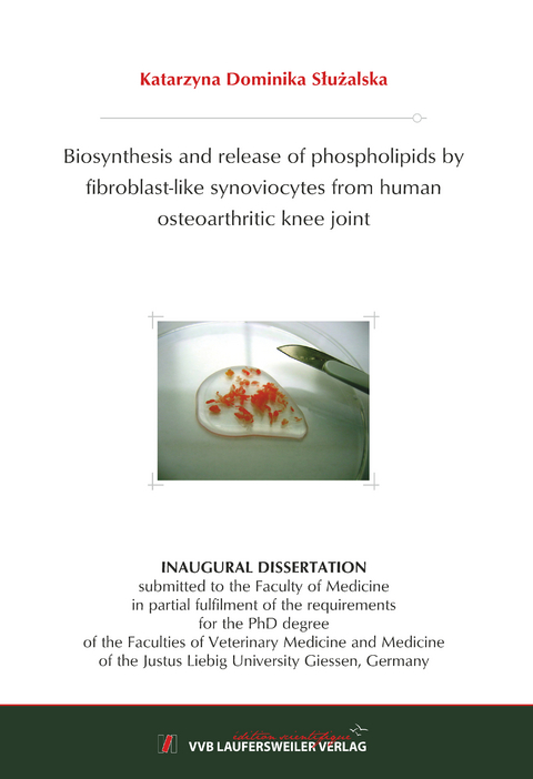
Biosynthesis and release of phospholipids by fibroblast-like synoviocytes from human osteoarthritic knee joint
Seiten
2017
VVB Laufersweiler Verlag
978-3-8359-6621-5 (ISBN)
VVB Laufersweiler Verlag
978-3-8359-6621-5 (ISBN)
- Keine Verlagsinformationen verfügbar
- Artikel merken
In human joints phospholipids (PLs) are produced and released by fibroblast-like synoviocytes (FLS). However, the regulatory mechanism of these processes remains poorly understood. Elevated levels of cytokines and growth factors as well as of PLs were found in synovial fluid (SF) during osteoarthritis (OA). Therefore, we hypothesized that PL metabolism in FLS is regulated by various agents being present in OA SF. This study aimed to develop two in vitro models to study the biosynthesis and release of PLs in order to evaluate the effects of cytokines, growth factors and drugs on PL metabolism.
To measure the biosynthesis of PLs, FLS were cultured in DMEM containing 5% lipoprotein deficient serum in the presence of stable isotope-labelled precursors of PLs and various agents. To study the release of PLs, FLS were cultured in DMEM containing 10% FBS in the presence of radiolabelled precursors of PLs. Cells were starved and the release of radiolabelled PLs was determined in DMEM containing 2% FBS in the presence of agents to be tested. Lipids were extracted from cellular lysates and media, and then quantified using electrospray ionization tandem mass spectrometry in the biosynthesis model or liquid scintillation counting in the release model.
The results of our lipidomic study provide for the first time a detailed overview of PLs being synthesized and released from human FLS. We were able to demonstrate that IL-1β induced the biosynthesis of phosphatidylethanolamine (PE) and PE-based plasmalogens (PE P), whereas TNFα induced only the biosynthesis of PE. Also, BMPs induced the biosynthesis of several PE and PE P species. In vivo PE P could protect against cartilage destruction mediated by ROS, whereas elevated PE could induce apoptosis of hypertrophic FLS and osteophytes. Furthermore, growth factors such as TGF-β1, IGF-1, and BMP-2 upregulated the biosynthesis of phosphatidylcholine (PC) which in vivo could be responsible for joint lubrication as well as mediation of the signal transduction. Additionally, dexamethasone was found to decrease the biosynthesis of PE. Moreover, we demonstrated that the release of radiolabelled PLs is a time-dependent process. However, tested agents did not influence the release of PLs using our in vitro model. Thus, the mechanism controlling PL release needs to be further investigated.
In conclusion, our results indicate that cytokines and growth factors regulate PL biosynthesis and may contribute to the altered PL composition in OA SF. Moreover, our data suggest that FLS undergo PL alterations to adapt to the new diseased environment. Understanding intra- and extracellular functions of elevated PLs within human articular joints is a new challenge for lipidomic studies.
| Erscheinungsdatum | 06.11.2017 |
|---|---|
| Reihe/Serie | Edition Scientifique |
| Sprache | englisch |
| Maße | 146 x 210 mm |
| Gewicht | 236 g |
| Themenwelt | Veterinärmedizin |
| Schlagworte | Doktorarbeit • Uni • Wissenschaft |
| ISBN-10 | 3-8359-6621-9 / 3835966219 |
| ISBN-13 | 978-3-8359-6621-5 / 9783835966215 |
| Zustand | Neuware |
| Informationen gemäß Produktsicherheitsverordnung (GPSR) | |
| Haben Sie eine Frage zum Produkt? |
Mehr entdecken
aus dem Bereich
aus dem Bereich
A Practical Guide
Buch | Hardcover (2024)
Wiley-Blackwell (Verlag)
145,95 €
Buch | Hardcover (2024)
Wiley-Blackwell (Verlag)
124,55 €
Buch | Softcover (2024)
Wiley-Blackwell (Verlag)
95,12 €


