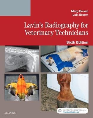
Lavin's Radiography for Veterinary Technicians
Elsevier - Health Sciences Division (Verlag)
978-0-323-41367-1 (ISBN)
- Titel erscheint in neuer Auflage
- Artikel merken
More than 1000 full-color photos and updated radiographic images visually demonstrate the relationship between anatomy and positioning.
UNIQUE! Non-manual restraint techniques including sandbags, tape, rope, sponges, sedation and combinations improve your safety and radiation protection.
UNIQUE! Comprehensive dental radiography coverage gives you a meaningful background in the dentistry subsection of vet radiography.
Increased emphasis on digital radiography, including quality factors and post-processing, keeps you up-to-date on the most recent developments in digital technology.
Broad coverage of radiologic science, physics, imaging and protection provide you with foundations for good technique.
Objectives, key terms, outlines, chapter introductions and key points help you organize information to ensure you understand what is most important in every chapter.
Color anatomy art created by an expert medical illustrator help you to recognize and avoid making imaging mistakes.
Check It Out boxes provide suggestions for practical actions that help better understand content being presented.
Points to ponder boxes emphasize information critical to performing tasks correctly.
Key points boxes help you to review critical content presented in the radiographic positioning chapters.
NEW! All chapters have been reviewed, revised and updated to present content in a way that is easy to follow and understand.
NEW! Updated radiation protection chapter focuses on the importance of safety in the lab.
NEW! Additional popular diagnostic information includes MRI/PET and CT/PET scans.
NEW! Coverage of Sante's Rule that clearly explains the mathematical process for creating a technique chart
NEW! Chapters on Dental Imaging and Radiography, Quality Control, and Testing and Artifacts combines existing content with updates into these important parts of radiography.
Marg Brown, RVT, BEd Ad Ed has been a veterinary technician educator for more than 30 years. Most of the time was spent at Seneca College of Applied Arts and Technology in Ontario as a full-time instructor. She also had the position of co-coordinator for several years. She received her Animal Health Technician diploma from Centralia College, a Bachelor of Education in Adult Education from Brock University and is a Registered Veterinary Technician. She has been involved in a variety of subjects over the years and was instrumental in revamping the core program at Seneca College. She is currently an adjunct faculty with Penn Foster in the Veterinary Technician program.
Part One:
Section 1: The Technical Side of Imaging 1. The Basics of Atoms and Electricity 2. Diagnostic Xray Production 3. Radiation Safety and Protection Section 2: Film and Digital Imaging 4. Imaging on Film 5. Producing the Image 6. Optimizing the Image 7. Processing the Image on Film 8. Computerized Radiography; Digital Imaging 9. Quality Control, Testing and Artifacts Section 3: Specialized Imaging 10. Ultrasound 11. Fluoroscopy 12. Computerized Tomography 13. Magnetic Resonance Imaging 14. Nuclear Medicine and Intro to P.E.T. Part Two: 15. Overview of Positioning 16. Small Animal Abdomen 17. Small Animal Thorax 18. Small Animal Forelimb 19. Small Animal Pelvis and Pelvic Limb 20. Small Animal Vertebral Column 21. Small Animal Skull 22. Dental Imaging and Radiography 23. Small Animal Special Procedures 24. Equine and Large Animal Radiography 25. Avian and Exotic Radiography
| Erscheinungsdatum | 17.12.2017 |
|---|---|
| Zusatzinfo | Approx. 1065 illustrations (1065 in full color); Illustrations |
| Verlagsort | Philadelphia |
| Sprache | englisch |
| Maße | 216 x 276 mm |
| Gewicht | 1290 g |
| Themenwelt | Veterinärmedizin ► Klinische Fächer ► Bildgebende Verfahren |
| ISBN-10 | 0-323-41367-6 / 0323413676 |
| ISBN-13 | 978-0-323-41367-1 / 9780323413671 |
| Zustand | Neuware |
| Haben Sie eine Frage zum Produkt? |
aus dem Bereich


