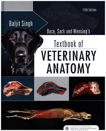
Dyce, Sack, and Wensing's Textbook of Veterinary Anatomy
Saunders (Verlag)
978-0-323-44264-0 (ISBN)
Gain the working anatomic knowledge that is crucial to your understanding of the veterinary basic sciences with Textbook of Veterinary Anatomy, 5th Edition. By focusing on the essential anatomy of each species, this well-established book details information directly applicable to the care of dogs, cats, horses, cows, pigs, sheep, goats, birds, and camelids - and points out similarities and differences among species. Each chapter includes a conceptual overview that describes the structure and function of an anatomic region, and new diagrams facilitate comprehension of bodily functions.
Comprehensive coverage of a multitude of species (dogs, cats, birds, horses, cows, pigs, sheep, goats, and now camelids) provides comparative anatomic information all in one resource.
Focuses on essential anatomy of each species, making this text ideal for any veterinary school curriculum that is covering the same amount of material in a reduced number of hours for course completion.
Clinical cases throughout relate actual cases to the study of anatomy to increase your understanding of how patients will present in practice.
Evolve site for students and instructors with a test bank, sample flash cards, and an image collection reinforces your understanding of veterinary anatomy - and helps you prepare for the NAVLE board exam.
Content is logically organized into two main sections - a general introduction to mammalian anatomy and a region-specific breakdown - to make studying more efficient and ensure greater understanding.
Flash cards and coloring book support the text and give you the opportunity to study anatomy in different ways to increase retention of what you have learned.
NEW and EXPANDED! New and updated diagrams further illustrate the anatomic structure and function of each species to facilitate comprehension of bodily functions, presenting the closest thing possible to an actual "hands-on" clinical experience.
EXPANDED! A 500-question Test Bank, designed for optimal NAVLE board exam preparation, lets you practice answering board-type questions.
UPDATED! New information on the anatomy of camelids, a species that is gaining importance in farm animal practice, provides an overview of the camelid anatomy and highlights any major differences compared to other species.
Baljit Singh , Vice-President, Research and Professor of Veterinary Anatomy, University of Saskatchewan, Saskatoon, Canada.
Part 1: General Anatomy
1. Some Basic Facts and Concepts
2. The Locomotor Apparatus
3. The Digestive Apparatus
4. The Respiratory Apparatus
5. The Urogenital Apparatus
6. The Endocrine Glands
7. The Cardiovascular System
8. The Nervous System
9. The Sense Organs
10. The Common Integument
Part 2: Dogs and Cats
11. The Head and Ventral Neck of the Dog and Cat
12. The Neck, Back, and Vertebral Column of the Dog and Cat
13. The Thorax of the Dog and Cat
14. The Abdomen of the Dog and Cat
15. The Pelvis and Reproductive Organs of the Dog and Cat
16. The Forelimb of the Dog and Cat
17. The Hindlimb of the Dog and Cat
Part 3: Horses
18. The Head and Ventral Neck of the Horse
19. The Neck, Back and Vertebral Column of the Horse
20. The Thorax of the Horse
21. The Abdomen of the Horse
22. The Pelvis and Reproductive Organs of the Horse
23. The Forelimb of the Horse
24. The Hindlimb of the Horse
Part 4: Ruminants
25. The Head and Ventral Neck of the Ruminant
26. The Neck, Back, and Vertebral Column of the Ruminant
27. The Thorax of the Ruminant
28. The Abdomen of the Ruminant
29. The Pelvis and Reproductive Organs of the Ruminant
30. The Forelimb of the Ruminant
31. The Hindlimb of the Ruminant
Part 5: Pigs
32. The Head and Neck of the Pig
33. The Vertebral Column, Back, and Thorax of the Pig
34. The Abdomen of the Pig
35. The Pelvis and Reproductive Organs of the Pig
36. The Limbs of the Pig
Part 6: Birds and Camelids
37. Anatomy of the Bird
38. Clinical Anatomy of Llamas and Alpacas
| Erscheinungsdatum | 10.07.2017 |
|---|---|
| Zusatzinfo | Approx. 1540 illustrations (1500 in full color); Illustrations |
| Verlagsort | Philadelphia |
| Sprache | englisch |
| Maße | 216 x 276 mm |
| Gewicht | 2400 g |
| Themenwelt | Veterinärmedizin ► Vorklinik ► Anatomie |
| Veterinärmedizin ► Kleintier | |
| Veterinärmedizin ► Großtier | |
| ISBN-10 | 0-323-44264-1 / 0323442641 |
| ISBN-13 | 978-0-323-44264-0 / 9780323442640 |
| Zustand | Neuware |
| Informationen gemäß Produktsicherheitsverordnung (GPSR) | |
| Haben Sie eine Frage zum Produkt? |
aus dem Bereich


