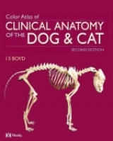
A Colour Atlas of Clinical Anatomy of the Dog and Cat
Seiten
1991
Mosby (Verlag)
978-0-7234-1649-4 (ISBN)
Mosby (Verlag)
978-0-7234-1649-4 (ISBN)
- Titel ist leider vergriffen;
keine Neuauflage - Artikel merken
Zu diesem Artikel existiert eine Nachauflage
Focuses on the anatomical study of the cat and the dog, with step-by-step "layering" of dissections and providing guidance on surgical approaches. Special emphasis is placed on regions of clinical interest throughout.
The study of anatomy is the fundamental basis of all teaching in the medical sciences. For many students, however, the connection between theory and practice is lost because of the amount of unnecessary information they are required to absorb. "A Colour Atlas of Small Animal Applied Anatomy" provides students and practitioners with a guide to the anatomy of the dog and cat which is relevant to the demands of small animal practice. While selective in its coverage, special emphasis is placed on those areas of clinical interest. Each section begins with an overview of the animal's surface anatomy, identifying key topographic landmarks of underlying anatomical features. The collection of photographs are of fresh, unfixed material, and demonstrate the normal appearance of the tissues. These are supplemented by radiographs, ultrasonographs, endoscopic views and line diagrams to aid interpretation of the more detailed dissections, providing the reader with a comprehensive picture of the body structure. Additional illustrations show the position and orientation of these structures in situ to aid the practitioner in planning surgical approaches.
The study of anatomy is the fundamental basis of all teaching in the medical sciences. For many students, however, the connection between theory and practice is lost because of the amount of unnecessary information they are required to absorb. "A Colour Atlas of Small Animal Applied Anatomy" provides students and practitioners with a guide to the anatomy of the dog and cat which is relevant to the demands of small animal practice. While selective in its coverage, special emphasis is placed on those areas of clinical interest. Each section begins with an overview of the animal's surface anatomy, identifying key topographic landmarks of underlying anatomical features. The collection of photographs are of fresh, unfixed material, and demonstrate the normal appearance of the tissues. These are supplemented by radiographs, ultrasonographs, endoscopic views and line diagrams to aid interpretation of the more detailed dissections, providing the reader with a comprehensive picture of the body structure. Additional illustrations show the position and orientation of these structures in situ to aid the practitioner in planning surgical approaches.
Head and neck; spinal column; pectoral limb; thorax; abdomen; pelvis; pelvic limb.
| Zusatzinfo | 90 line diagrams, 75 b&w and 254 colour photographs, index |
|---|---|
| Verlagsort | London |
| Sprache | englisch |
| Maße | 254 x 308 mm |
| Gewicht | 1346 g |
| Themenwelt | Veterinärmedizin ► Vorklinik ► Anatomie |
| Veterinärmedizin ► Kleintier | |
| ISBN-10 | 0-7234-1649-4 / 0723416494 |
| ISBN-13 | 978-0-7234-1649-4 / 9780723416494 |
| Zustand | Neuware |
| Informationen gemäß Produktsicherheitsverordnung (GPSR) | |
| Haben Sie eine Frage zum Produkt? |
Mehr entdecken
aus dem Bereich
aus dem Bereich
Buch | Softcover (2024)
Lehmanns Media (Verlag)
39,95 €
Lehrbuch und Farbatlas für Studium und Praxis
Buch | Hardcover (2024)
Thieme (Verlag)
195,00 €


