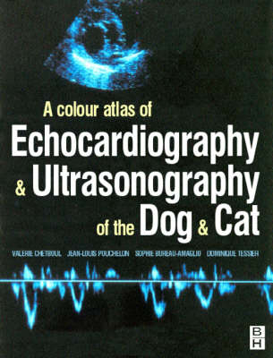
Echocardiography and Ultrasound of the Dog and Cat
Butterworth-Heinemann Ltd (Verlag)
978-0-7506-4856-1 (ISBN)
- Titel wird leider nicht erscheinen
- Artikel merken
Ultrasonography is a very new technique for veterinarians. This practical and unique four-colour atlas firstly gives an overview of the physical principles of acquiring an echocardiograph and Doppler image, then discusses the different stages of the examination. It contains 2-D and 3-D diagrams which show how to carry out an examination and how to interpret the final image in terms of the pathology according to the position of the sound in the animal's thorax and the image obtained in the screen. Not only will it be of use to students but it will also be an invaluable source of reference to those practising veterinarians who wish to discover the potentials of using such equipment before a final investment.
Chapter 1 - Physical Principles of Echography and Doppler Echo Echocardiographic imaging : general principles Basic properties of ultrasounds Physical properties of ultrasounds Interaction of ultrasounds with tissues Transducers Piezo-electric crystals Intrinsic parameters of transducers Different types of probes and screening techniques Generating echographic images Patient preparation Positioning the animal Choosing the right probe Image quality Doppler examination of the heart : general principles The Doppler effect: physical principles and analysis Physical principles of the Doppler effect Analysis of the Doppler effect Different Doppler modes Continuous wave Doppler Pulsed wave Doppler Colour coded Doppler Chapter 2 - Echocardiography and Doppler echo: physiological data The normal echocardiographic examination Bi-dimensional mode Definition Principal anatomical sections Time-Motion mode Definition Principal anatomical sections Main Indexes Echocardiographic normal values Inter-ventricular septal motion (IVS) in TM mode The normal Doppler examination Atrio-ventricular flows Mitral flow Tricuspid flow Arterial flows Aortic flow Pulmonic flow Chapter 3 - Echocardiography and Doppler examination of the diseased heart Congenital Cardiopathies Valvular abnormalities Arterial stenosis and incompetence Atrio-ventricular valve dysplasia Shunts Patent ductus arteriosus (PDA) Ventricular septal defects (VSD) Atrial septal defects (ASD) Atrio-ventricular canal Pericardial abnorm
| Übersetzer | Martin du Breuil |
|---|---|
| Zusatzinfo | colour illustrations, index |
| Verlagsort | London |
| Sprache | englisch |
| Maße | 189 x 246 mm |
| Themenwelt | Veterinärmedizin ► Klinische Fächer ► Bildgebende Verfahren |
| Veterinärmedizin ► Kleintier | |
| ISBN-10 | 0-7506-4856-2 / 0750648562 |
| ISBN-13 | 978-0-7506-4856-1 / 9780750648561 |
| Zustand | Neuware |
| Haben Sie eine Frage zum Produkt? |
aus dem Bereich


