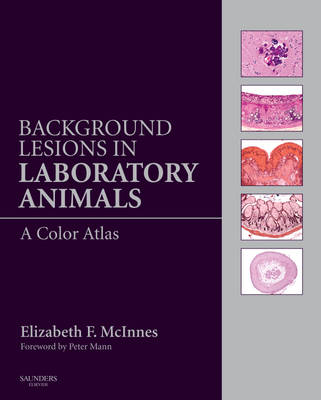
Background Lesions in Laboratory Animals
A Color Atlas
Seiten
2011
W B Saunders Co Ltd (Verlag)
978-0-7020-3519-7 (ISBN)
W B Saunders Co Ltd (Verlag)
978-0-7020-3519-7 (ISBN)
- Titel ist leider vergriffen;
keine Neuauflage - Artikel merken
Suitable for pathologists needing to recognize background and incidental lesions while examining slides taken from laboratory animals in acute and chronic toxicity studies, or while examining exotic species in a diagnostic laboratory, this title gives them descriptions and illustrations of majority of background lesions likely to be encountered.
Background Lesions in Laboratory Animals will be an invaluable aid to pathologists needing to recognize background and incidental lesions while examining slides taken from laboratory animals in acute and chronic toxicity studies, or while examining exotic species in a diagnostic laboratory. It gives clear descriptions and illustrations of the majority of background lesions likely to be encountered. Many of the lesions covered are unusual and can be mistaken for treatment-related findings in preclinical toxicity studies.
The Atlas has been prepared with contributions from experienced toxicological pathologists who are specialists in each of the laboratory animal species covered and who have published extensively in these areas.
over 600 high-definition, top-quality color photographs of background lesions found in rats, mice, dogs, minipigs, non-human primates, hamsters, guinea pigs and rabbits
a separate chapter on lesions in the reproductive systems of all laboratory animals written by Dr Dianne Creasy, a world expert on testicular lesions in laboratory animals
a chapter on common artifacts that may be observed in histological glass slides
extensive references to each lesion described
aging lesions encountered in all laboratory animal species, particularly in rats in mice which are used for carcinogenicity studies
Chapter 1 - Non human primates. Cynomolgus monkey (Maccaca fascicularis) and marmoset (Callithrix jacchus)
Chapter 2 - Wistar and CD rat
Chapter 3 - Beagle Dog
Chapter 4 - Mouse
Chapter 5 - Syrian Hamster
Chapter 6 - Minipig
Chapter 7 - Rabbit
Chapter 8 - Artifacts in processed tissues
Chapter 9 - Reproduction of the Rat, Dog, Primate and Pig
| Erscheint lt. Verlag | 11.10.2011 |
|---|---|
| Vorwort | Peter Mann |
| Zusatzinfo | Approx. 623 illustrations (623 in full color) |
| Verlagsort | London |
| Sprache | englisch |
| Maße | 276 x 219 mm |
| Themenwelt | Medizin / Pharmazie ► Medizinische Fachgebiete ► Notfallmedizin |
| Studium ► 2. Studienabschnitt (Klinik) ► Pathologie | |
| Veterinärmedizin ► Vorklinik | |
| Veterinärmedizin ► Kleintier | |
| ISBN-10 | 0-7020-3519-X / 070203519X |
| ISBN-13 | 978-0-7020-3519-7 / 9780702035197 |
| Zustand | Neuware |
| Haben Sie eine Frage zum Produkt? |
Mehr entdecken
aus dem Bereich
aus dem Bereich


