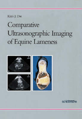
Comparative Ultrasonographic Imaging of Equine Lameness
Seiten
- Titel ist leider vergriffen;
keine Neuauflage - Artikel merken
Ultrasonography has become a very important imaging modality in the diagnosis and management of equine lameness. The selections of normal and abnormal ultrasonographic images and the correspondending text offered by this book demonstrates the usefullness of ultrasound for non-invasive imaging of various tendons and ligaments, muscles, articular and peri-articular soft tissue structures, distended tendons sheaths and bursa of the front and hind limb. For better understanding of the ultrasound images, photographs of cross sectional sections of frozen limb secimen and computed tomographic images are included. Additional survey and contrast radiographic studies illustrate the complmentary value and limitations of these procedures with ultrasongraphic imaging.
| Zusatzinfo | 291 Abb., davon 30 farb. |
|---|---|
| Sprache | englisch |
| Maße | 343 x 245 mm |
| Gewicht | 1080 g |
| Themenwelt | Veterinärmedizin ► Klinische Fächer ► Pathologie |
| Schlagworte | HC/Medizin/Veterinärmedizin • Pferd • Pferde; Veterinärmedizin • Tierheilkunde • Ultraschalldiagnostik • Ultraschalldiagnostik (vet.) • Ultraschalldiagnostik (Veterinärmedizin) |
| ISBN-10 | 3-87706-523-6 / 3877065236 |
| ISBN-13 | 978-3-87706-523-5 / 9783877065235 |
| Zustand | Neuware |
| Haben Sie eine Frage zum Produkt? |
Mehr entdecken
aus dem Bereich
aus dem Bereich
Atlas der morphologischen Zellbestimmung
Buch | Softcover (2021)
LABOKLIN GmbH & Co. KG (Verlag)
55,95 €


