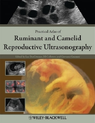
Practical Atlas of Ruminant and Camelid Reproductive Ultrasonography
Iowa State University Press (Verlag)
978-0-8138-1551-0 (ISBN)
Editor-in-chief: Luc DesCoteaux, DMV, MSc, Dipl. ABVP (Dairy), is a Full Professor and Service Chief of the Ambulatory Clinic at the Universite de Montreal, Quebec, Canada. Associate Editors: Giovanni Gnemmi, DVM, PhD, Dipl. ECBHM, is a bovine practitioner and ultrasound instructor with BovineVet Italy. Jill Colloton, DVM, is a bovine practitioner and ultrasound instructor with Bovine Services, LLC, USA.
Preface. Acknowledgments. Introduction. Chapter 1 Principles and recommendations, essential concepts, and common artifacts in ultrasound imaging. Description and practical recommendations in the choice of ultrasound equipment with a view to image quality. General principles and essential concepts to improve image quality. Common artifacts. Chapter 2 Scanning techniques and common errors in bovine practice. Description of scanning technique. Manipulation of the probe. Common errors. Chapter 3 Anatomy of the reproductive tract of the cow. Genital tract. Descriptive terminology of the ovary and ovarian structures. Chapter 4 Bovine ovary. Endocrinology and ovarian structures in pubertal cows. Ovarian anomalies and differential diagnosis. Use of color Doppler to monitor blood flow. Ultrasound use in reproduction synchronization protocols for dairy cattle: Two perspectives. Chapter 5 Bovine uterus. Ultrasound of the uterus during the estrous cycle and normal postpartum period. Color Doppler sonography of the uterine blood flow. Ultrasound of the postpartum abnormal uterus and vagina. Chapter 6 Bovine pregnancy. Morphologic embryonic and fetal development up to day 55. Ultrasound landmarks of standard early pregnancy diagnosis. Early embryonic and fetal death. Twins. Chapter 7 Bovine fetal development after 55 days, fetal sexing, anomalies, and well-being. Fetal development after 55 days. Ultrasound fetal sexing. Fetal anomalies. Fetal well-being during late pregnancy (normal gestation, compromised pregnancy, and clone). Chapter 8 Bovine embryo transfer, in vitro fertilization, special procedures, and cloning. Embryo donors. Oocyte collection for in vitro fertilization. Recipients. Management of clone recipients. Chapter 9 Bull anatomy and ultrasonography of the reproductive tract. Ultrasound equipment and techniques. Anatomy of the reproductive system. Anomalies and ultrasonographic imaging of external and internal reproductive organs. Chapter 10 Buffalo and zebu cattle. Equipment and scanning techniques. Major differences between bovine and bubaline species. Pathology. Congenital and hereditary defects. Ultrasound services in buffalo and zebu. Chapter 11 Sheep and goats. Usefulness of ultrasonography in small ruminants. Equipment and scanning techniques. Ultrasound imaging of the female reproductive tract. Endocrine and ovarian processes that comprise the normal estrous cycle and pregnancy. Fetal count, age, and sex. Pathological conditions in the female. Ultrasonographic evaluation of the male genital system. Common abnormalities of the testis. Common abnormalities of the accessory glands. Chapter 12 Camelids. Usefulness of ultrasonography in camelids. Equipment and scanning techniques. Ultrasonographic anatomy. Ovarian function and endocrinology in South American camelids. Pregnancy diagnosis and evaluation of fetal growth. Uterine and ovarian abnormalities. Index.
| Erscheint lt. Verlag | 12.1.2010 |
|---|---|
| Verlagsort | Arnes, AI |
| Sprache | englisch |
| Maße | 224 x 286 mm |
| Gewicht | 1142 g |
| Themenwelt | Veterinärmedizin ► Klinische Fächer ► Bildgebende Verfahren |
| Veterinärmedizin ► Klinische Fächer ► Gynäkologie / Geburtshilfe | |
| Veterinärmedizin ► Klinische Fächer ► Pathologie | |
| Veterinärmedizin ► Großtier ► Krankheitslehre | |
| ISBN-10 | 0-8138-1551-7 / 0813815517 |
| ISBN-13 | 978-0-8138-1551-0 / 9780813815510 |
| Zustand | Neuware |
| Informationen gemäß Produktsicherheitsverordnung (GPSR) | |
| Haben Sie eine Frage zum Produkt? |
aus dem Bereich


