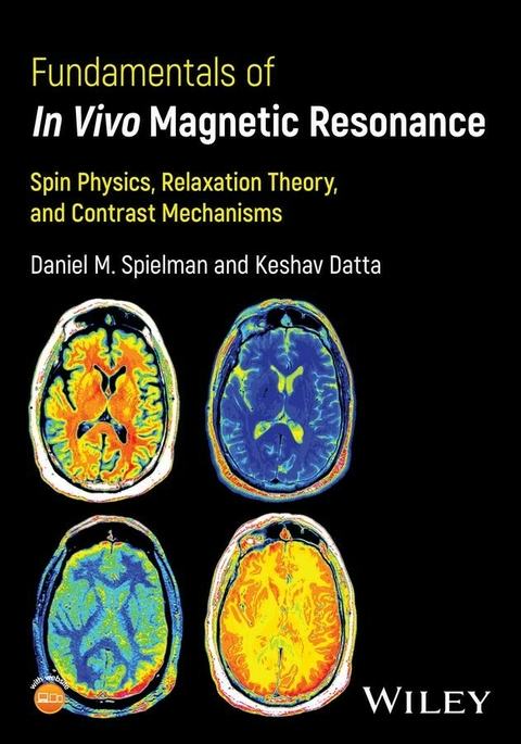
Fundamentals of In Vivo Magnetic Resonance
John Wiley & Sons Inc (Verlag)
978-1-394-23309-0 (ISBN)
Daniel M. Spielman, PhD, is Professor of Radiology at Stanford University, Stanford, CA, USA. He is a fellow of both the American Institute for Medical & Biological Engineering (AIMBE) and International Society of Magnetic Resonance in Medicine (ISMRM), and has received multiple teaching awards including the ISMRM Outstanding Teacher Award (2005) and Stanford Department of Radiology Research Faculty of the Year (2022).
Keshav Datta, PhD, is Vice President, Research & Development, at VIDA Diagnostics Inc., Coralville, IA, USA, a precision lung health company, accelerating therapies to patients through AI-powered lung intelligence.. He is also a Consulting Research Scientist at Stanford University, Stanford, CA, USA.
Chapter 1. Introduction
1.1. A Brief History of MR
1.2. NMR vs MRI
1.3. The Roadmap
1.4. Historical Notes
Chapter 2. Classical Description of MR
2.1. Nuclear Magnetism
2.2. Net Magnetization and the Bloch Equations
2.3. Rf Excitation and Reception
2.4. Spatial Localization
2.5. The MRI Signal Equation
2.6. Exercises
2.7. Historical Notes
Chapter 3. Quantum Description of MR
3.1. Introduction
3.1.1. Why QM for magnetic resonance?
3.1.2. Historical developments
3.1.3. Wavefunctions
3.2. Mathematics of QM
3.2.1. Linear vector spaces
3.2.2. Dirac notation and Hilbert Space
3.2.3. Liouville Space
3.3. The Six Postulates of QM
3.4. MR in Hilbert Space
3.4.1. Review of spin operators
3.4.2. Single spin in a magnetic field: longitudinal and transverse magnetization
3.4.3. Ensemble of spins in a magnetic field
3.5. MR in Liouville Space
3.5.1. Statistical mixture of quantum states
3.5.2. The density operator
3.5.3. The Spin-lattice Disconnect
3.5.4. Hilbert space vs Liouville space
3.5.5. Observations about the spin density operator
3.5.6. Solving the Liouville-von Neuman equation
3.6. Exercises
3.7. Historical Notes
Chapter 4. Nuclear Spins
4.1. Review of the Spin Density Operator and the Hamiltonian
4.2. External Interactions
4.3. Internal Interactions
4.3.1. Chemical shift
4.3.2. Dipolar coupling
4.3.3. J-coupling
4.4. Summary of the Nuclear Spin Hamiltonian
4.5. Exercises
4.6. Historical Notes
Chapter 5. Product Operator Formulism
5.1. The Density Operator, Populations, and Coherences
5.1.1. Spin systems and associated density operators
5.1.2. Density matrix calculations
5.2. POF for Single-Spin Coherence Space
5.3. POF for Two-Spin Coherence Space
5.4. Branch Diagrams
5.5. Multiple Quantum Coherences and 2D NMR
5.6. Polarization Transfer
5.7. Spectral Editing
5.7.1. J-difference editing
5.7.2. Multiple quantum filtering
5.8. Exercises
5.9. Historical Notes
Chapter 6. In vivo MRS
6.1. 1H MRS
6.1.1. Acquisition methods
6.1.2. Detectable metabolites and applications
6.2. 31P-MRS
6.3. 13C-MRS
6.3.1. Acquisition methods
6.3.2. 13C infusion studies
6.3.3. Hyperpolarized 13C
6.4. Deuterium Metabolic Imaging
6.5. 23Na-MRI
6.6. Exercises
Chapter 7. Relaxation Fundamentals
7.1. Basic Principles
7.1.1. Molecular motion
7.1.2. Stochastic processes
7.1.3. A simple model of relaxation
7.2. Dipolar Coupling
7.2.1. The Solomon equations
7.2.2. Calculating transition rates
7.2.3. Nuclear Overhauser Effect
7.3. Chemical Exchange
7.3.1. Introduction
7.3.2. Effects on longitudinal magnetization
7.3.3. Effects on transverse magnetization
7.3.4. Examples
7.4. In vivo Water
7.4.1. Hydration layers
7.4.2. Tissue relaxation times
7.4.3. Magic angle effects
7.4.4. Magnetization Transfer Contrast (MTC)
7.4.5. Chemical Exchange Saturation Transfer (CEST)
7.5. Exercises
7.6. Historical Notes
Chapter 8. Redfield Theory of Relaxation
8.1. Perturbation theory and the Interaction Frame of Reference
8.2. Calculating Relaxation Times
8.3. Relaxation mechanisms
8.3.1. Dipolar coupling revisited
8.3.2. Scalar relaxation of the 1st kind and 2nd kind
8.3.3. Chemical Shift Anisotropy (CSA)
8.4. Relaxation in the Rotating Frame
8.4.1. Physics of T1r
8.4.2. The spin-lock experiment
8.4.3. Applications
8.5. Illustrative Examples
8.5.1. Hyperpolarized 13C-urea
8.5.2. Hyperpolarized 13C-Pyr
8.6. Exercises
8.7. Historical Notes
Chapter 9. MRI Contrast Agents
9.1. Paramagnetic Relaxation Enhancement
9.1.1. Solomon-Bloembergen-Morgan theory
9.1.2. Gd3+-based T1 contrast agents
9.2. T2 and T2* Contrast Agents
9.2.1. T2, diffusion, and outer-sphere relaxation
9.2.2. SPIOs and USPIOs
9.3. PARACEST Contrast Agents
9.4. Contrast Agents in the Clinic
9.4.1. Gd-based agents
9.4.2. Iron-based agents
9.5. Exercises
Chapter 10. In vivo Examples
10.1. Relaxation properties of brain
10.1.1. Morphological imaging
10.1.2. Perfusion imaging
10.1.3. Diffusion-Weighted Imaging (DWI)
10.1.4. Imaging myelin
10.1.5. Susceptibility-Weighted Imaging (SWI)
10.2. Relaxation properties of blood
10.2.1. Hemoglobin and red blood cells
10.2.2. MRI blood oximetry
10.2.3. functional Magnetic Resonance Imaging (fMRI)
10.2.4. MRI of hemorrhage
10.3. Relaxation properties of cartilage
10.3.1. T2 mapping
10.3.2. DWI
10.3.3. T1r mapping and dispersion
10.3.4. gagCEST
10.3.5. dGEMRIC
10.3.6. Ultrashort TE (UTE) imaging
10.3.7. Sodium MRI
10.3.8. Summary
10.4. Synopsis
10.5. Exercises
Exercise Solutions
References
Index
| Erscheinungsdatum | 21.03.2024 |
|---|---|
| Verlagsort | New York |
| Sprache | englisch |
| Gewicht | 798 g |
| Einbandart | kartoniert |
| Themenwelt | Medizinische Fachgebiete ► Radiologie / Bildgebende Verfahren ► Kernspintomographie (MRT) |
| Naturwissenschaften ► Physik / Astronomie ► Angewandte Physik | |
| Technik ► Maschinenbau | |
| Technik ► Medizintechnik | |
| ISBN-10 | 1-394-23309-4 / 1394233094 |
| ISBN-13 | 978-1-394-23309-0 / 9781394233090 |
| Zustand | Neuware |
| Informationen gemäß Produktsicherheitsverordnung (GPSR) | |
| Haben Sie eine Frage zum Produkt? |
aus dem Bereich


