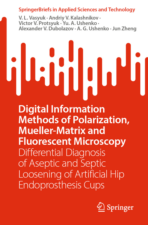
Digital Information Methods of Polarization, Mueller-Matrix and Fluorescent Microscopy
Springer Verlag, Singapore
978-981-99-4734-8 (ISBN)
This book highlights the effectiveness of differential diagnosis in the degree of severity of joint pathology from a clinical, biophysical, and informational point of view. It includes the following information blocks:
- Two-dimensional digital polarization microscopy of polycrystalline films of synovial fluid and determination of the coordinate distributions of the orientation and phase parameters of the microscopic image from a set of parameters of the Stokes vector.
- Mueller-matrix mapping of polycrystalline films of synovial fluid and determination of a set of coordinate distributions (Mueller-matrix images (MMI)) of azimuthal-invariant elements that characterize manifestations of optical activity and linear birefringence.- Development of algorithms for polarization reproduction of distributions of linear and circular birefringence of polycrystalline films of synovial fluid.
- Identification of digital statistical, correlation and wavelet criteria of polarization and Mueller-matrix differential diagnosis of the degree of severity of joint pathology.
- Determination of maps of laser-induced fluorescence of synovial fluid polycrystalline films.
- Identification of statistical and correlational criteria for fluorescent differential diagnosis of the degree of severity of joint pathology.
- Operational characteristics of the power of the methods of azimuth-invariant polarization, Mueller-matrix and laser autofluorescence microscopy of polycrystalline films of synovial fluid.
Dr. Vasyuk V.L.- Orthopedic surgeon of the highest category, Ph. D., Doctor of medicine, professor, Head of the Department of Traumatology and Orthopedics at Bukovinian State Medical University, Supervisor at Regional center "Traumatology, endoprosthetics, joint and spine pathology". Author of 372 publications, 6 monographs, 220 articles, 25 Patents of USSR, 8 Patents of Ukraine. Dr. Kalashnikov A.V. – MD, professor, head of the department of Trauma Injuries to Locomotion System and Problems of Osteosynthesis at the SI “The Institute of Traumatology and Orthopedics, NAMS of Ukraine”, Dr. Protsyuk Victor V.- Orthopedic surgeon of the highest category, Ph. D., Supervisor at Regional center "Traumatology, endoprosthetics, joint and spine pathology". Dr. Yuriy A. Ushenko obtained his Ph.D. in optics and laser physics in 2006, and D.Sc. in optics and laser physics from the Taras Shevchenko National University of Kyiv in 2015. He is currently Professor and Head of the Computer Science Department at Chernivtsi National University, Ukraine. His current research interests include laser polarimetry, holography, and application of AI in life sciences. Dr. Alexander Dubolazov is an Associate Professor at the Chernivtsi National University. He received his BS and MS degrees in optics from the Chernivtsi National University in 2006 and 2007, respectively, and his PhD degree in optics, laser physics from the Chernivtsi National University in 2010. He is the author of more than 100 journal papers and has written five book chapters. His current research interests include laser polarimetry, holography etc. Dr. Alexander G. Ushenko is Head of Optics and Publishing Department at the Chernivtsi National University, Ukraine. He received his BS and MS degrees in optics from the Chernivtsi State University in 1975 and 1977, respectively, and his Ph.D. degree inoptics and laser physics from the Chernivtsi State University in 1983. In 2000, he obtained D.Sc. in optics and laser physics. His current research interests include laser polarimetry, polarization interferometry, and digital holography. Dr. Zheng Jun is an associate research fellow in Research Institute of Zhejiang University-Taizhou. He obtained the Ph.D. degree in Control Theory and Control Engineering from Zhejiang University, China in 2005. He has been engaged in the application research of industrial automation system and its advanced control, photoelectric detection and intelligent perception for a long time. In recent years, Zheng Jun has presided over two major cooperation research projects with China Railway Construction Electrification Bureau Group and participated in two national key research and development projects.
lt;p>Introduction
1. Methods and Means of Polarization, Mueller-Matrix, Polarization-Correlation and Fluorescence Diagnostics in Medicine
Abstract
1.1. Methods and Means of Polarization and Mueller-Matrix Microscopy of Biological Objects
1.2. Azimuthal-Invariant Mueller-Matrix Microscopy of Optically Anisotropic Polycrystalline Films of Human Organs
1.3. Methods for Fluorescence Diagnostics of Biological Structures
References2. Materials and Optical-Physical and Fluorescent Research Methods
Abstract2.1. Design and Structural-Logical Diagram of The Study of the Polycrystalline Structure of SF Films
2.2. Model Structure of Polycrystalline Films of Synovial Fluid
2.3. Structural-Logical Diagram and Techniques of Polarization and Mueller-Matrix Microscopy of Synovial Fluid Layers2.4. Structural-Logical Diagram and Techniques of Spectral-Selective Laser-Induced
Fluorescence Microscopy of Polycrystalline Films of Synovial Fluid
2.5. Analytical algorithms for data processing
2.5.1. Statistical analysis
2.5.2. Information analysis
2.6. Brief description of research objects2.7. Conclusions to Chapter 2
References
3. Computer Digital Differential Diagnostics of Aseptic and Septic Loosening of Endoprosthesis Cup of Artificial Hip Joint by Methods of Polarizing Microscopy
Abstract
3.1. Structural and logical diagram of polarizing microscopy
3.2. Differential Diagnosis of Aseptic and Septic Loosening of the Endoprosthesis Cup of an Artificial Hip Joint by Polarization Microscopy of the Distributions of the Orientation Parameter of Digital Images of Polycrystalline Films of Synovial Fluid
3.2.1. Information Analysis of the Data of the Method of Mapping the Distributions of the Value of Digital Microscopic Images of Polycrystalline Films of SF
3.3. Differential Diagnosis of Aseptic and Septic Loosening of the Cup of an Artificial Hip Joint Endoprosthesis by the Method of Polarization Microscopy of the Phase Parameter Distributions of Digital Images of Polycrystalline Films of the Synovial Fluid of the Knee Joint
3.3.1. Information Analysis of the Data of the Method of Mapping the Distributions of the Value of PP of Digital Microscopic Images of Polycrystalline Films of SF
3.4. Wavelet Analysis of OP Maps of Microscopic Images of Polycrystalline Films of SF of the Hip Joint in Patients With Varying Severity of Pathology
3.4.1. Informational Analysis of Wavelet Analysis Data of The Method of Mapping the Distributions of the OP Value of Digital Microscopic Images of Polycrystalline Films of SF
3.5. Wavelet Analysis of PP Maps of Microscopic Images of Polycrystalline Films of SF of The Hip Joint in Patients with Varying Severity of Pathology
3.5.1. Informational Analysis of Wavelet Analysis Data of The Method of Mapping the Distributions of The PP Value of Digital Microscopic Images of Polycrystalline Films of SF
3.6. Conclusions to Chapter 3References
4. Differential Diagnostics of Aseptic and Septic Loosening of the Artificial Hip Joint Endoprosthesis Cup by Methods of Azimuthal-Invariant Mueller-Matrix Microscopy
Abstract
4.1. Structural and Logical Diagram of Azimuthal-Invariant Mueller-Matrix Microscopy
4.2. Differential Diagnosis of Aseptic and Septic Loosening of The Cup of An Artificial Hip Joint Endoprosthesis by the Method of Azimuthally Invariant Mueller-Matrix Microscopy
of the MMI Distributions of Optical Activity of Polycrystalline Films of the Synovial Fluid of the Hip Joint
4.2.1. Informational Analysis of the Data of the Method of Mapping the Distributions
of the MMI OA Value for Polycrystalline Films of SF4.3. Differential Diagnosis of Aseptic and Septic Loosening of the Cup of an Artificial Hip Joint Endoprosthesis by the Method of Mueller-Matrix Microscopy of the Distributions of the MMI LB Value of Polycrystalline Films of Synovial Fluid
4.3.1. Informational Analysis of the Data of the Method of Mapping the Distributions of the PP Value of Digital Microscopic Images of Polycrystalline Films Of SF4.4. Wavelet Analysis of MMI OA Maps of Polycrystalline Films of SF of Hip Joint in Patients
with Varying Severity of Pathology4.4.1 Informational Analysis of Wavelet Analysis Data of the Method of Mapping the Distributions of the MMI OA Value for Polycrystalline Films of SF
4.5. Wavelet Analysis of MMI LB Maps of Polycrystalline Films of Hip Joint SF In Patients
with Varying Severity of Pathology
4.5.1. Informational Analysis of the Wavelet Analysis Data of the Mueller-Matrix Mapping of the Distributions of the MMI LB Value for Polycrystalline Films of SF
4.6. Conclusions to Chapter 4References
5. Polarization Tomography Methods and Differential Diagnostics Aseptic and Septic Loosening of the Artificial Hip Joint Endoprosthesis Cup
Abstract
5.1. Structural and Logical Diagram of Differential Mueller-Matrix Tomography
of The Polycrystalline Structure of SF Films
5.2. Differential Diagnosis of Aseptic and Septic Loosening of the Cup of an Artificial Hip Joint Endoprosthesis by the Mueller-Matrix Reconstruction of the Distributions of CB Of Polycrystalline Films of The Synovial Fluid of the Hip Joint
5.2.1. Information Analysis of the Data of the Mueller-Matrix Reconstruction of the Distributions of the Circular Birefringence of Polycrystalline SF Films
5.3. Differential Diagnosis of Aseptic and Septic Loosening of the Cup of an Artificial Hip Joint Endoprosthesis by the Method of Mueller-Matrix Reconstruction of the Distributions
of the LB Value of Polycrystalline Films of Synovial Fluid of the Hip Joint
5.3.1. Information Analysis of the Data of the Mueller-Matrix Reconstruction of the Distributions of the Linear Birefringence of Polycrystalline Films of SF5.4. Wavelet Analysis of CB Maps of Polycrystalline Films of SF of the Hip Joint in Patients
with Varying Severity of Pathology5.4.1. Informational Analysis of Wavelet Analysis Data on the Distributions of the Value of CB of Polycrystalline Films of SF
5.5. Wavelet Analysis of LB Maps of Polycrystalline Films of SF of the Hip in Patients
with Varying Severity of Pathology
5.5.1. Informational Analysis of Wavelet-Analysis Data of the Mueller-Matrix Reproduction of the Distributions of the LB Value of Polycrystalline Films of SF
5.6. Conclusions to Chapter 5References
6. Differential Diagnosis of The Aseptic and Septic Loosening of the Artificial Hip Joint Endoprosthesis Methods of Spectral-Selective Laser Autofluorescence Microscopy
Abstract
6.1. Structural and Logical Diagram of Spectral-Selective Laser Autofluorescence Microscopy
of Maps of Coordinate Distribution (MIF) and Correlation (MCF) of the Fluorescence Intensity of Polycrystalline Films of SF of the Hip Joint
6.2. Differential Diagnosis of Aseptic and Septic Loosening of the Cup of an Artificial Hip Joint Endoprosthesis by Spectral-Selective Fluorescence Microscopy of the Coordinate Distributions of the Autofluorescence Intensity of Polycrystalline Films of
Synovial Fluid
6.3. Informational Analysis of Spectral-Selective Fluorescence Microscopy Datafor Polycrystalline Films of SL
6.4. Differential Diagnosis of Aseptic and Septic Loosening of the Cup of an Artificial Hip Joint Endoprosthesis by the Method of Correlation Analysis of Spectral-Selective MIF of Polycrystalline Films of Synovial Fluid
6.5. Informational Analysis of Data from the Method of Correlation Analysis of Spectral-Selective Maps of Fluorescence Intensity of Polycrystalline Films of SF
6.6. Conclusions to Chapter 6
References 7.0 Results and Conclusions| Erscheinungsdatum | 24.08.2023 |
|---|---|
| Reihe/Serie | SpringerBriefs in Applied Sciences and Technology |
| Zusatzinfo | 48 Illustrations, color; 10 Illustrations, black and white; XIV, 102 p. 58 illus., 48 illus. in color. |
| Verlagsort | Singapore |
| Sprache | englisch |
| Maße | 155 x 235 mm |
| Themenwelt | Naturwissenschaften ► Chemie ► Analytische Chemie |
| Naturwissenschaften ► Physik / Astronomie ► Optik | |
| Technik ► Elektrotechnik / Energietechnik | |
| Schlagworte | Autofluorescence Microscopy • Joint pathology • Laser Images • Mueller-Matrix Microscopy • Numerical Methods • Polarizing Microscopy • Polycrystalline Films • synovial fluid |
| ISBN-10 | 981-99-4734-0 / 9819947340 |
| ISBN-13 | 978-981-99-4734-8 / 9789819947348 |
| Zustand | Neuware |
| Haben Sie eine Frage zum Produkt? |
aus dem Bereich


