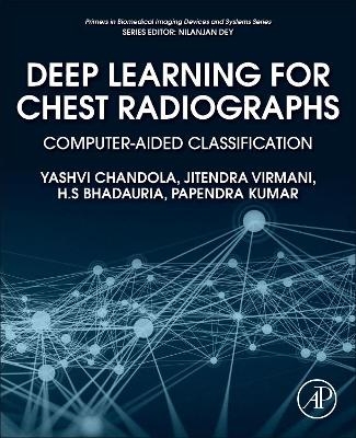
Deep Learning for Chest Radiographs
Academic Press Inc (Verlag)
978-0-323-90184-0 (ISBN)
This book is a valuable resource for academicians, researchers, clinicians, postgraduate and graduate students in medical imaging, CAC, computer-aided diagnosis, computer science and engineering, electrical and electronics engineering, biomedical engineering, bioinformatics, bioengineering, and professionals from the IT industry.
Yashvi Chandola received her B-Tech (Hons.) in Computer Science and Engineering from Women Institute of Technology, Dehradun, Uttarakhand in 2018. She has completed her M-Tech (Hons.) in Computer Science and Engineering from Govind Ballabh Pant Institute of Engineering and Technology, Pauri Garhwal, Uttarakhand in 2020. Her research interests include application of machine learning and deep learning algorithms for analysis of medical images. Jitendra Virmani received his B-Tech (Hons.) in Instrumentation Engineering from Sant Longowal Institute of Engineering and Technology, Punjab in 1999 and M-Tech in Electrical Engineering with specialization in Measurement and Instrumentation from the Indian Institute of Technology, Roorkee in 2006. He served in the academia for nine years before joining the PhD programme in 2009 at Biomedical Instrumentation Laboratory, Electrical Engineering Department, Indian Institute of Technology, Roorkee as a full time Research Scholar under MHRD Assistantship. He received his PhD from the Indian Institute of Technology, Roorkee in 2013. After his PhD he served in Academia for Jaypee University of Information Technology, Solan, Himachal Pradesh and Thapar Institute of Engineering and Technology, Patiala, Punjab before joining CSIR-CSIO, Chandigarh in August 2016. He is presently working at CSIR-CSIO, Chandigarh. He is a Life member of the Institute of Engineers (IEI), India and Computer Society of India. He has published 85 papers in various journals, conferences and Book chapters with various reputed publishers. He has delivered more than 35 expert talks on various platforms basically on application of machine learning and deep learning algorithms for medical images. He is Editorial Board Member of International Journal of Image Mining published by Inderscience Publishers. His research interests include application of machine learning and deep learning algorithms for analysis of medical images. H.S Bhadauria received his B-Tech in Computer Science and Engineering from Aligarh Muslim University, Aligarh in 1999, and M-Tech in Electronics Engineering from Aligarh Muslim University, Aligarh in 2004. He received his PhD on Detection and Segmentation of Brain Hemorrhage using CT images from Biomedical Signal and Image Processing Laboratory, Indian Institute of Technology - Roorkee in 2013. During his PhD he worked on enhancing the detection and segmentation of brain hemorrhage using CT imaging modality. He has served in academia for more than 12 years. He is presently serving as a Professor in the Department of Computer Science & Engineering at Govind Ballabh Pant Institute of Engineering and Technology, Pauri Garhwal, Uttarakhand. He is a life member of Institute of Engineers (IEI), India. He has published more than 60 research papers in International and National Journals and Conferences. His areas of research interest are Digital Image and Digital Signal Processing. Papendra Kumar received his B.E in Computer Science and Engineering from Govind Ballabh Pant Institute of Engineering and Technology, Pauri Garhwal, Uttarakhand in 2007 and M-Tech in Digital Signal Processing from Govind Ballabh Pant Institute of Engineering and Technology, Pauri Garhwal, Uttarakhand in 2011. He is presently serving as an Assistant Professor in the Department of Computer Science & Engineering at Govind Ballabh Pant Institute of Engineering and Technology, Pauri Garhwal, Uttarakhand. His areas of research interest are Digital Image and Digital Signal Processing.
1. Introduction 2. Review of Related Work 3. Methodology Adopted for Designing of Computer-Aided Classification Systems for Chest Radiographs 4. End-to-end Pre-trained CNN-based Computer-Aided Classification System design for Chest Radiographs 5. Hybrid Computer-Aided Classification System Design Using End-to-end Pre-trained CNN-based Deep Feature Extraction and ANFC-LH Classifier for Chest Radiographs 6. Hybrid Computer-Aided Classification System Design Using End-to-end Pre-trained CNN-based Deep Feature Extraction and PCA-SVM Classifier for Chest Radiographs 7. Light-weight End-to-end Pre-trained CNN-based Computer-Aided Classification System Design for Chest Radiographs 8. Hybrid Computer-Aided Classification System Design Using Light-weight End-to-end Pre-trained CNN-based Deep Feature Extraction and ANFC-LH Classifier for Chest Radiographs 9. Hybrid Computer-Aided Classification System Design Using Light-weight End-to-end Pre-trained CNN-based Deep Feature Extraction and PCA-SVM Classifier for Chest Radiographs 10. Comparative Analysis of Computer-Aided Classification Systems Designed for Chest Radiographs: Conclusion and Future Scope
| Erscheinungsdatum | 30.07.2021 |
|---|---|
| Reihe/Serie | Primers in Biomedical Imaging Devices and Systems |
| Zusatzinfo | 60 illustrations (20 in full color); Illustrations |
| Verlagsort | Oxford |
| Sprache | englisch |
| Maße | 191 x 235 mm |
| Gewicht | 500 g |
| Themenwelt | Informatik ► Theorie / Studium ► Künstliche Intelligenz / Robotik |
| Informatik ► Weitere Themen ► Bioinformatik | |
| Medizin / Pharmazie ► Medizinische Fachgebiete ► Radiologie / Bildgebende Verfahren | |
| Technik | |
| ISBN-10 | 0-323-90184-0 / 0323901840 |
| ISBN-13 | 978-0-323-90184-0 / 9780323901840 |
| Zustand | Neuware |
| Haben Sie eine Frage zum Produkt? |
aus dem Bereich


