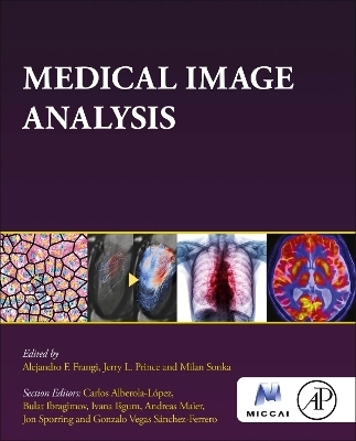
Medical Image Analysis
Academic Press Inc (Verlag)
978-0-12-813657-7 (ISBN)
Alejandro F. Frangi is the Bicentennial Turing Chair in Computational Medicine and Royal Academy of Engineering Chair in Emerging Technologies at The University of Manchester, Manchester, UK, with joint appointments at the Schools of Engineering (Department of Computer Science), Faculty of Science and Engineering, and the School of Health Sciences (Division of Informatics, Imaging and Data Science), Faculty of Biology, Medicine and Health. He is a Turing Fellow of the Alan Turing Institute. He holds an Honorary Chair at KU Leuven in the Departments of Electrical Engineering (ESAT) and Cardiovascular Science. He is IEEE Fellow (2014), EAMBES Fellow (2015), SPIE Fellow (2020), MICCAI Fellow (2021), and Royal Academy of Engineering Fellow (2023). The IEEE Engineering in Medicine and Biology Society awarded him the Early Career Award (2006) and Technical Achievement Award (2021). Professor Frangi’s primary research interests are in medical image analysis and modeling, emphasising machine learning (phenomenological models) and computational physiology (mechanistic models). He is an expert in statistical shape modeling, computational anatomy, and image-based computational physiology, delivering novel insights and impact across various imaging modalities and diseases, particularly on cardiovascular MRI, cerebrovascular MRI/CT/3DRA, and musculoskeletal CT/DXA. He is a co-founder of adsilico Ltd., and his work led to products commercialized by GalgoMedical SA. He has published over 285 peer-reviewed papers in scientific journals with over 34,000 citations and has an h-index of 75. Jerry L. Prince is the William B. Kouwenhoven Professor in the Department of Electrical and Computer Engineering at Johns Hopkins University. He is Director of the Image Analysis and Communications Laboratory (IACL). He also holds joint appointments in the Departments of Radiology and Radiological Science, Biomedical Engineering, Computer Scienceand Applied Mathematics and Statistics at Johns Hopkins University. He received a 1993 National Science Foundation Presidential Faculty Fellows Award, was Maryland’s 1997 Outstanding Young Engineer, and was awarded the MICCAI Society Enduring Impact Award in 2012. He is an IEEE Fellow, MICCAI Fellow, and AIMBE Fellow. Previously he was an Associate Editor of IEEE Transactions on Image Processing and an Associate Editor of IEEE Transactions on Medical Imaging. He is currently a member of the Editorial Boards of Medical Image Analysis and the Proceedings of the IEEE. He is cofounder of Sonavex, Inc., a biotech company located in Baltimore, Maryland, USA. His current research interests include image processing, computer vision, and machine learning with primary application to medical imaging, he has published over 500 articles on these subjects. Milan Sonka is Professor of Electrical & Computer Engineering, Biomedical Engineering, Ophthalmology & Visual Sciences, and Radiation Oncology, and Lowell C. Battershell Chair in Biomedical Imaging, all at the University of Iowa. He served as Chair of the Department of Electrical and Computer Engineering (2008–2014) and as Associate Dean for Research and Graduate Studies (2014–2019). He is a Fellow of IEEE, Fellow of the American Institute of Medical and Biological Engineers (AIMBE), Fellow of the Medical Image Computing and Computer-Aided Intervention Society (MICCAI), and Fellow of the National Academy of Inventors. He is the Founding Codirector of an interdisciplinary Iowa Institute for Biomedical Imaging (2007–) and Founding Director of the Iowa Initiative for Artificial Intelligence (2019–). He is the author of four editions of an image processing textbook, Image Processing, Analysis, and Machine Vision (1993, 1998, 2008, 2014), editor of one of three volumes of the SPIE Handbook of Medical Imaging (2000), past Editor-in-Chief of “IEEE Transactions on Medical Imaging (2009–2014), and past editorial board member of the “Medical Image Analysis journal. His >700 publications were cited more than 42,000 times, and he has an h-index of 80. He cofounded Medical Imaging Applications LLC and VIDA Diagnostics Inc.
PART I Introductory topics
1. Medical imaging modalities
2. Mathematical preliminaries
3. Regression and classification
4. Estimation and inference
PART II Image representation and processing
5. Image representation and 2D signal processing
6. Image filtering: enhancement and restoration
7. Multiscale and multiresolution analysis
PART III Medical image segmentation
8. Statistical shape models
9. Segmentation by deformable models
10. Graph cut-based segmentation
PART IV Medical image registration
11. Points and surface registration
12. Graph matching and registration
13. Parametric volumetric registration
14. Non-parametric volumetric registration
15. Image mosaicking
PART V Machine learning in medical image analysis
16. Deep learning fundamentals
17. Deep learning for vision and representation learning
18. Deep learning medical image segmentation
19. Machine learning in image registration
PART VI Advanced topics in medical image analysis
20. Motion and deformation recovery and analysis
21. Imaging Genetics
PART VII Large-scale databases
22. Detection and quantitative enumeration of objects from large images
23. Image retrieval in big image data
PART VIII Evaluation in medical image analysis
24. Assessment of image computing methods
| Erscheinungsdatum | 30.09.2023 |
|---|---|
| Reihe/Serie | The MICCAI Society book Series |
| Verlagsort | San Diego |
| Sprache | englisch |
| Maße | 191 x 235 mm |
| Gewicht | 1000 g |
| Themenwelt | Medizin / Pharmazie ► Medizinische Fachgebiete ► Radiologie / Bildgebende Verfahren |
| Medizin / Pharmazie ► Physiotherapie / Ergotherapie ► Orthopädie | |
| Technik ► Medizintechnik | |
| ISBN-10 | 0-12-813657-X / 012813657X |
| ISBN-13 | 978-0-12-813657-7 / 9780128136577 |
| Zustand | Neuware |
| Informationen gemäß Produktsicherheitsverordnung (GPSR) | |
| Haben Sie eine Frage zum Produkt? |
aus dem Bereich


