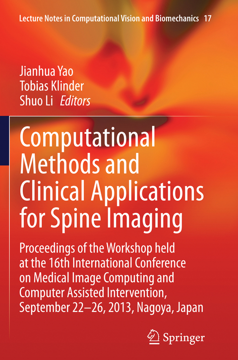
Computational Methods and Clinical Applications for Spine Imaging
Springer International Publishing (Verlag)
978-3-319-35805-5 (ISBN)
Preface.- Workshop Organization.- Segmentation I (CT): Segmentation of vertebrae from 3D spine images by applying concepts from transportation and game theories, by Bulat Ibragimov, Bostjan Likar, Franjo Pernus, Tomaz Vrtovec.- Automatic and Reliable Segmentation of Spinal Canals in Low-Resolution, Low-Contrast CT Images, by Qian Wang, Le Lu, Diji Wu, Noha El-Zehiry, Dinggang Shen, Kevin Zhou.- A Robust Segmentation Framework for Spine Trauma Diagnosis, by Poay Hoon Lim, Ulas Bagci, Li Bai.- 2D-PCA based Tensor Level Set Framework for Vertebral Body Segmentation, by Ahmed Shalaby, Aly Farag, Melih Aslan.- Computer Aided Detection and Diagnosis: Computer Aided Detection of Spinal Degenerative Osteophytes on Sodium Fluoride PET/CT, by Jianhua Yao, Hector Munoz , Joseph Burns, Le Lu, Ronald Summers.- Novel Morphological and Appearance Features for Predicting Physical Disability from MR Images in Multiple Sclerosis Patients, by Jeremy Kawahara, Chris McIntosh, Roger Tam, Ghassan Hamarneh.- Classification of Spinal Deformities using a Parametric Torsion Estimator, by Jesse Shen, Stefan Parent, Samuel Kadoury.- Lumbar Spine Disc Herniation Diagnosis with a Joint Shape Model, by Raja Alomari, Vipin Chaudhary, Jason Corso, Gurmeet Dhillon.- Epidural Masses Detection on Computed Tomography Using Spatially-Constrained Gaussian Mixture Models, by Sanket Pattanaik, Jiamin Liu, Jianhua Yao, Weidong Zhang, Evrim Turkbey, Xiao Zhang, Ronald Summers.- Quantitative Imaging: Comparison of manual and computerized measurements of sagittal vertebral inclination in MR images, by Tomaz Vrtovec, Franjo Pernus, Bostjan Likar.- Eigenspine: Eigenvector Analysis of Spinal Deformities in Idiopathic Scoliosis, by Daniel Forsberg, Claes Lundström, Mats Andersson, Hans Knutsson.- Quantitative Monitoring of Syndesmophyte Growth in Ankylosing Spondylitis Using Computed Tomography, by Sovira Tan, Jianhua Yao,Lawrence Yao, Michael Ward.- A Semi-automatic Method for the Quantification of Spinal Cord Atrophy, by Simon Pezold, Michael Amann, Katrin Weier, Ketut Fundana, Ernst Radue, Till Sprenger, Philippe Cattin.- Segmentation II (MR): Multi-modal vertebra segmentation from MR Dixon in hybrid whole-body PET/MR, by Christian Buerger, Jochen Peters, Irina Waechter-Stehle, Frank Weber, Tobias Klinder, Steffen Renisch.- Segmentation of intervertebral discs from high-resolution 3D MRI using multi-level statistical shape models, by Ales Neubert, Jurgen Fripp, Craig Engstrom, Stuart Crozier.- A supervised approach towards segmentation of clinical MRI for automatic lumbar diagnosis, by Subarna Ghosh, Manavender Malgireddy, Vipin Chaudhary, Gurmeet Dhillon.- Registration/Labeling: Automatic Segmentation and Discrimination of Connected Joint Bones from CT by Multi-atlas Registration, by Tristan Whitmarsh, Graham Treece, Kenneth Poole.- Registration of MR to Percutaneous Ultrasound of the Spine for Image-Guided Surgery, by Lars Eirik Bø, Rafael Palomar, Tormod Selbekk, Ingerid Reinertsen.- Vertebrae Detection and Labelling in Lumbar MR Images, by Meelis Lootus, Timor Kadir, Andrew Zisserman.
| Erscheinungsdatum | 15.09.2016 |
|---|---|
| Reihe/Serie | Lecture Notes in Computational Vision and Biomechanics |
| Zusatzinfo | XI, 230 p. 92 illus. |
| Verlagsort | Cham |
| Sprache | englisch |
| Maße | 155 x 235 mm |
| Themenwelt | Informatik ► Grafik / Design ► Digitale Bildverarbeitung |
| Informatik ► Theorie / Studium ► Künstliche Intelligenz / Robotik | |
| Medizinische Fachgebiete ► Radiologie / Bildgebende Verfahren ► Radiologie | |
| Medizin / Pharmazie ► Physiotherapie / Ergotherapie ► Orthopädie | |
| Technik | |
| Schlagworte | biomedical engineering • Computer Imaging, Vision, Pattern Recognition and • computer vision • Engineering • Engineering: general • Image Based Modeling • Image Processing • Image Segmentation • Imaging / Radiology • Medical Imaging • MICCAI • MICCAI 2013 • Radiology • spine imaging |
| ISBN-10 | 3-319-35805-7 / 3319358057 |
| ISBN-13 | 978-3-319-35805-5 / 9783319358055 |
| Zustand | Neuware |
| Informationen gemäß Produktsicherheitsverordnung (GPSR) | |
| Haben Sie eine Frage zum Produkt? |
aus dem Bereich


