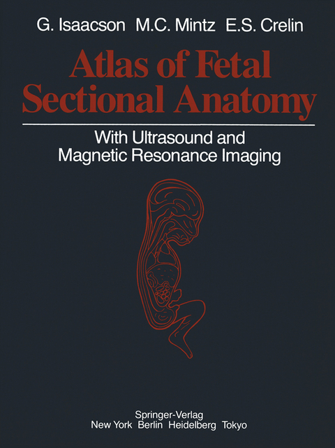
Atlas of Fetal Sectional Anatomy
With Ultrasound and Magnetic Resonance Imaging
Seiten
2013
|
Softcover reprint of the original 1st ed. 1986
Springer-Verlag New York Inc.
978-1-4613-8617-9 (ISBN)
Springer-Verlag New York Inc.
978-1-4613-8617-9 (ISBN)
The fetal period of human growth and development has become an area of intense study in recent years, due in large part to the development of diagnostic ultrasound. These points include the changing anatomy of the fetal brain during gestation and the anatomy of the meninges, the fetal heart, and ductus venosus.
The fetal period of human growth and development has become an area of intense study in recent years, due in large part to the development of diagnostic ultrasound. More than 2,000 articles have been published in the last five years describing anatomy and pathology in utero, as reflected in sonographic images. Yet, no stan dard reference exists to correlate these images with fetal gross anatomy and at tempts to draw parallels from adult structure have often led to false assumptions. The dictum "the newborn is not a miniature adult" is all the more valid for the fetus. This text aims to provide a comprehensive reference for normal sectional anat omy correlated with in utero ultrasound images. In addition, magnetic resonance images of therapeutically aborted or stillborn fetuses are paired with similar gross sections to serve as a foundation upon which current in vivo studies may build. Lastly, a miscellaneous section illustrates several anatomic points useful in the understanding of fetal anatomy. These points include the changing anatomy of the fetal brain during gestation and the anatomy of the meninges, the fetal heart, and ductus venosus. It is our hope that this atlas will provide a clear picture of fetal anatomy, rectify some of the confusion which exists in antenatal diagnosis, and stimulate further interest in fetal development.
The fetal period of human growth and development has become an area of intense study in recent years, due in large part to the development of diagnostic ultrasound. More than 2,000 articles have been published in the last five years describing anatomy and pathology in utero, as reflected in sonographic images. Yet, no stan dard reference exists to correlate these images with fetal gross anatomy and at tempts to draw parallels from adult structure have often led to false assumptions. The dictum "the newborn is not a miniature adult" is all the more valid for the fetus. This text aims to provide a comprehensive reference for normal sectional anat omy correlated with in utero ultrasound images. In addition, magnetic resonance images of therapeutically aborted or stillborn fetuses are paired with similar gross sections to serve as a foundation upon which current in vivo studies may build. Lastly, a miscellaneous section illustrates several anatomic points useful in the understanding of fetal anatomy. These points include the changing anatomy of the fetal brain during gestation and the anatomy of the meninges, the fetal heart, and ductus venosus. It is our hope that this atlas will provide a clear picture of fetal anatomy, rectify some of the confusion which exists in antenatal diagnosis, and stimulate further interest in fetal development.
Fetal Head and Neck.- 1 Sagittal Sections.- 2 Coronal Sections.- 3 Axial Sections.- Fetal Thorax and Abdomen.- 1 Axial Sections.- Male Thorax and Abdomen.- Male Pelvis.- Female Pelvis.- 2 Sagittal Sections.- 3 Coronal Sections.- Fetal Limbs.- Special Studies.- 1 Fetal Brain Development.- Cortical Development.- Ventricular Development.- Dural Structures.- 2 Anatomy of the Ductus Venosus.- 3 Fetal Heart.
| Zusatzinfo | 253 Illustrations, black and white; XI, 184 p. 253 illus. |
|---|---|
| Verlagsort | New York, NY |
| Sprache | englisch |
| Maße | 210 x 280 mm |
| Themenwelt | Medizin / Pharmazie ► Gesundheitsfachberufe ► Hebamme / Entbindungspfleger |
| Medizin / Pharmazie ► Medizinische Fachgebiete ► Gynäkologie / Geburtshilfe | |
| Medizinische Fachgebiete ► Radiologie / Bildgebende Verfahren ► Sonographie / Echokardiographie | |
| Studium ► 1. Studienabschnitt (Vorklinik) ► Anatomie / Neuroanatomie | |
| Technik | |
| Schlagworte | anatomy • Ultrasound |
| ISBN-10 | 1-4613-8617-9 / 1461386179 |
| ISBN-13 | 978-1-4613-8617-9 / 9781461386179 |
| Zustand | Neuware |
| Haben Sie eine Frage zum Produkt? |
Mehr entdecken
aus dem Bereich
aus dem Bereich
Begleitbuch für Sonografiekurse, Klinik und Praxis
Buch | Softcover (2023)
Urban & Fischer in Elsevier (Verlag)
27,00 €
Organbezogene Darstellung von Grund- und Aufbaukurs sowie …
Buch | Hardcover (2020)
Deutscher Ärzteverlag
99,99 €


