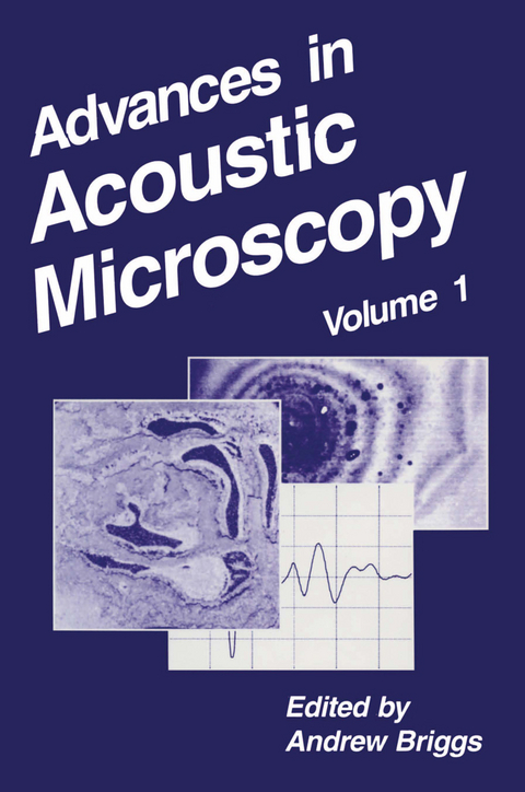
Advances in Acoustic Microscopy
Springer-Verlag New York Inc.
978-1-4613-5762-9 (ISBN)
1. Acoustic Microscopy Analysis of Microelectronic Interconnection and Packaging Technologies.- 1.1. Introduction.- 1.2. Ceramic Packages.- 1.3. Plastic IC Packages.- 1.4. Die Attach.- 1.5. Multilayer Interconnect.- 1.6. Tape Automated Bonding.- 1.7. Mechanical Properties.- 1.8. Future Perspectives.- 1.9. Conclusions.- Acknowledgments.- References.- 2. Measuring Short Cracks by Time-Resolved Acoustic Microscopy.- 2.1. Introduction.- 2.2. Short-Pulse System for the SAM.- 2.3. Time-Resolved Measurement Technique.- 2.4. Crack Depth Measurements in Plastic Material.- 2.5. Applying TOFD to Metals.- 2.6. Crack Growth Measurement.- 2.7. Discussion.- 2.8. Summary.- Acknowledgments.- References.- 3. Probing Biological Cells and Tissues with Acoustic Microscopy.- 3.1. Introduction.- 3.2. Probing Cells in Culture.- 3.3. Probing Microspheres and Freshly Isolated Outer Hair Cells.- 3.4. Bone and Other Collagenous Tissues.- 3.5. Model Investigations of Gels and Membranes.- 3.6. Conclusions.- Acknowledgments.- References.- 4. Lens Geometries for Quantitative Acoustic Microscopy.- 4.1. Introduction.- 4.2. Acoustic Lens Geometries.- 4.3. Comparison of Signal Processing Electronics.- 4.4. The V(f) Characterization Method.- 4.5. Accuracy of Velocity Measurement Using the V(z) Method..- 4.6. Conclusions.- Acknowledgments.- References.- 5. Measuring Thin-Film Elastic Constants by Line-Focus Acoustic Microscopy.- 5.1. Overview.- 5.2. Introduction.- 5.3. Measurement Model for V(z) Curves.- 5.4. Measuring and Calculating SAW Velocities.- 5.5. Inverse Method.- 5.6. Elastic Constants of Single-Layer Films.- 5.7. Effective Elastic Constants of Superlattices.- 5.8. Conclusion.- Acknowledgment.- References.- 6. Measuring the Elastic Properties of Stressed Materials by Quantitative Acoustic Microscopy.- 6.1. Introduction.- 6.2. Acoustoelasticity and Surface Waves.- 6.3. Line-Focus Beam Acoustic Microscopy.- 6.4. Determining Materials Properties.- 6.5. Examples.- 6.6. Conclusions.- Acknowledgments.- References.- 7. Surface Brillouin Scattering—Extending Surface Wave Measurements to 20 GHz.- 7.1. Introduction.- 7.2. Kinematics of Brillouin Scattering.- 7.3. Theory of Brillouin-Scattering Cross Section.- 7.4. Surface Brillouin Scattering Setup.- 7.5. Experimental Results.- 7.6. Conclusions.- Acknowledgments.- References.- 8. New Approaches in Acoustic Microscopy for Noncontact.- Measurement and Ultrahigh Resolution.- 8.1. Introduction.- 8.2. Phase Velocity Scanning Method.- 8.3. Atomic Force Microscopy (AFM) with a Vibrating Sample.- Acknowledgement.- References.
| Reihe/Serie | Advances in Acoustic Microscopy ; 1 |
|---|---|
| Zusatzinfo | XXXI, 350 p. |
| Verlagsort | New York, NY |
| Sprache | englisch |
| Maße | 152 x 229 mm |
| Themenwelt | Naturwissenschaften ► Biologie ► Allgemeines / Lexika |
| Naturwissenschaften ► Chemie ► Analytische Chemie | |
| Naturwissenschaften ► Geowissenschaften ► Mineralogie / Paläontologie | |
| Naturwissenschaften ► Physik / Astronomie ► Angewandte Physik | |
| Naturwissenschaften ► Physik / Astronomie ► Atom- / Kern- / Molekularphysik | |
| Naturwissenschaften ► Physik / Astronomie ► Festkörperphysik | |
| Naturwissenschaften ► Physik / Astronomie ► Thermodynamik | |
| Technik ► Maschinenbau | |
| ISBN-10 | 1-4613-5762-4 / 1461357624 |
| ISBN-13 | 978-1-4613-5762-9 / 9781461357629 |
| Zustand | Neuware |
| Informationen gemäß Produktsicherheitsverordnung (GPSR) | |
| Haben Sie eine Frage zum Produkt? |
aus dem Bereich


