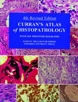
Curran's Atlas of Histopathology
Seiten
2000
|
4th Revised edition
Oxford University Press (Verlag)
978-0-19-263220-3 (ISBN)
Oxford University Press (Verlag)
978-0-19-263220-3 (ISBN)
- Titel ist leider vergriffen;
keine Neuauflage - Artikel merken
This fourth edition has been completely revised and there are additional immunohistological images. Most of the conditions are common or fairly common diseases, but occasional rare lesions are included. The book is primarily an atlas, the primary purpose of which is to convey information visually.
For this fourth edition of Professor Curran's colour atlas of histopathology, the text has been completely revised and there have been additional immunohistological images added to the 804 colour illustrations. The general arrangement of the contents has been retained, with a chapter on each of the main systems or organs of the body. There is an introductory chapter of a general nature which demonstrates the more important reactions of the tissues in disease and at the same time teaches the student the basic language of histopathology, thereby enabling him or her to read and assess the significance of changes in the tissue as revealed by microscopy. Most of the conditions are common or fairly common diseases, but occasional rare lesions are included. The book is primarily an atlas, the primary purpose of which is to convey information in visual form. It is meant to complement existing textbooks. A new comprehensive index has been prepared for this edition. This book is intended for primarily intended for undergraduate medical students but as with its predecessors, likely to prove useful to postgraduate students in training in pathology or other clinical disciplines.
For this fourth edition of Professor Curran's colour atlas of histopathology, the text has been completely revised and there have been additional immunohistological images added to the 804 colour illustrations. The general arrangement of the contents has been retained, with a chapter on each of the main systems or organs of the body. There is an introductory chapter of a general nature which demonstrates the more important reactions of the tissues in disease and at the same time teaches the student the basic language of histopathology, thereby enabling him or her to read and assess the significance of changes in the tissue as revealed by microscopy. Most of the conditions are common or fairly common diseases, but occasional rare lesions are included. The book is primarily an atlas, the primary purpose of which is to convey information in visual form. It is meant to complement existing textbooks. A new comprehensive index has been prepared for this edition. This book is intended for primarily intended for undergraduate medical students but as with its predecessors, likely to prove useful to postgraduate students in training in pathology or other clinical disciplines.
Preface; Methods; 1. General Pathology; 2. Blood, Spleen, Lymph Nodes and Bone Marrow; 3. Ear, Nose and Mouth; 4. Alimentary Tract; 5. Live, Gall Bladder and Pancreas; 6. Heart and Arteries; 7. Trachea, Bronchi and Lungs; 8. Endocrine Organs; 9. Central Nervous System and Eye; 10. Kidneys and Bladder; 11. Male Generative System; 12. Female generative System, including Breast; 13. Bones, Cartilage and Joints; 14. Skin; Index
| Erscheint lt. Verlag | 9.3.2000 |
|---|---|
| Zusatzinfo | 810 colour illustrations |
| Verlagsort | Oxford |
| Sprache | englisch |
| Maße | 210 x 276 mm |
| Gewicht | 1151 g |
| Themenwelt | Schulbuch / Wörterbuch ► Lexikon / Chroniken |
| Studium ► 1. Studienabschnitt (Vorklinik) ► Histologie / Embryologie | |
| Studium ► 2. Studienabschnitt (Klinik) ► Pathologie | |
| ISBN-10 | 0-19-263220-5 / 0192632205 |
| ISBN-13 | 978-0-19-263220-3 / 9780192632203 |
| Zustand | Neuware |
| Haben Sie eine Frage zum Produkt? |
Mehr entdecken
aus dem Bereich
aus dem Bereich
Zytologie, Histologie und mikroskopische Anatomie
Buch | Hardcover (2022)
Urban & Fischer in Elsevier (Verlag)
54,00 €


