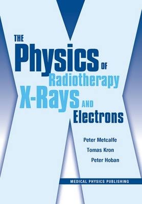
The Physics of Radiotherapy X-Rays and Electrons
Medical Physics Publishing Corporation (Verlag)
978-1-930524-36-1 (ISBN)
- Titel erscheint in neuer Auflage
- Artikel merken
This book is an updated successor to The Physics of Radiotherapy X-Rays from Linear Accelerators published in 1997. This new volume includes a significant amount of new material, including new chapters on electrons in radiotherapy and IMRT, IGRT, and tomotherapy, which have become key developments in radiation therapy.
Also updated from the earlier edition are the physics beam modeling chapters, including Monte Carlo methods, adding those mysterious electrons, as well as discourse on radiobiological modeling including TCP, NTCP, and EUD and the impact of these concepts on plan analysis and inverse planning.
This book is intended as a standard reference text for postgraduate radiation oncology medical physics students. It will also be of interest to radiation oncology registrars and residents, dosimetrists, and radiation therapists. The new text contains review questions at the end of each chapter and full bibliographic entries. Fully indexed. Selected questions and answers from The Q Book, The Physics of Radiotherapy X-Rays: Problems and Solutions are updated and integrated into the text.
Preface
Acknowledgments
Chapter 1-Medical Linear Accelerators
1.1 Introduction
1.2 Principles of Operation
1.3 Klystrons and Magnetrons
1.4 Electron Gun14
1.5 Accelerator Waveguide
1.6 Beam Delivery
1.7 Collimation
1.8 Wedges
1.9 Compensators
1.10 Electronic Portal Imaging Devices
1.11 On-Board Imaging Devices
1.12 Helical Tomotherapy
1.13 Gamma Knife and CyberKnifeReferences
Questions
Chapter 2-Interaction Properties of X-Rays and Electrons
2.1 Introduction
2.2 Cross Sections and Attenuation Coefficients
2.3 Photon Interaction Processes
2.4 Photon Beam Transport
2.5 Electron Interactions
2.6 Absorbed DoseReferences
Questions
Chapter 3-Dosimetry of Megavoltage X-Rays and Electrons
3.1 Introduction: Why Measure Dose?
3.2 The Ideal Dosimeter: Some Definitions
3.3 Radiotherapy Dosimetry Phantoms
3.4 Theory of Ionization Chamber Dosimetry
3.5 Ionization Chambers: Commercial Chambers and Electrometers
3.6 Semiconductor Detectors
3.7 Thermoluminescent Dosimeters
3.8 Film Dosimetry
3.9 Other Means of Dosimetry
3.10 Dosimeter Overview
References
Questions
Chapter 4-X-Ray Beam Properties
4.1 Introduction
4.2 Calibration Field Factors
4.3 Build-up Region
4.4 Percentage Depth Dose
4.5 Isocentric Dose Ratios
4.6 Beam Dose Profiles
4.7 Attenuation and Shielding
4.8 A Few Comments on kV X-Rays
References
Questions
Chapter 5-Linear Accelerator Electron Beam Properties
5.1 Introduction
5.2 Characterization of a Clinical Electron Beam
5.3 Depth Doses and Profiles
5.4 Electron Applicators and Cut-outs
5.5 Small and Irregular Fields
5.6 Variations in Geometry
5.7 Inhomogeneities
5.8 Typical Electron Beam Applications
5.9 A Few Comments on Protons
References
Questions
Chapter 6-Radiotherapy Treatment Planning: X-Rays
6.1 Introduction
6.2 Isodose Curves
6.3 Monitor Unit Calculations
6.4 Classical Simulation
6.5 Computed Tomography Simulation
6.6 Other Treatment Planning Aids
6.7 Radiotherapy Treatment Planning Hardware
6.8 Dose Display
6.9 Dose-Volume Histograms
6.10 Defining Treatment Planning Volumes
References
Questions
Chapter 7-Special Procedures: Stereotactic Radiotherapy and IMRT
7.1 The Rationale for Stereotactic Radiosurgery
7.2 Clinical Indications for SRS
7.3 Traditional Stereotactic Targeting
7.4 Frameless Stereotactic Targeting
7.5 SRS Treatment Planning
7.6 MMLC-based Radiosurgery Techniques
7.7 Robotic Radiosurgery: The CyberKnife
7.8 Stereotactic Body Radiotherapy
7.9 The Rationale for IMRT
7.10 Computation of Optimal Intensity Patterns for IMRT
7.11 MLC Leaf Sequences for IMRT
7.12 Dosimetric Properties of MLCs
7.13 Independent Plan Dosimetry Verification
7.14 Clinical Treatment with IMRT
7.15 Tomotherapy as an IMRT Modality
7.16 Analogy of Tomotherapy to CT Imaging
7.17 The TomoTherapy Hi-Art System®
7.18 Helical TomoTherapy Parameters
7.19 Clinical Use of Helical TomoTherapy
References
Questions
Chapter 8-Calibration of Megavoltage Photon and Electron Beams
8.1 Introduction
8.2 Absolute Dosimetry with Ionization Chambers
8.3 Air-Kerma Calibration-based Protocols
8.4 Dose-to-Water Calibration-based Protocols
8.5 Uncertainty Analysis and Verification
References
Questions
Chapter 9-Beam Models: Part I
9.1 Introduction
9.2 The Milan/Bentley Model
9.3 Inhomogeneity Corrections
9.4 Electron Pencil Beams
9.5 Brachytherapy Source Models
References
Questions
Chapter 10-Beam Models: Part II
10.1 Introduction
10.2 Monte Carlo Simulation
10.3 BEAM
10.4 Geant4
10.5 Convolutions/Superposition Methods
10.6 Macro Monte Carlo
References
Questions
Chapter 11-Quality Assurance in Radiotherapy
11.1 Introduction
11.2 Definition of Terms
11.3 Equipment That Requires QA
11.4 Intensity-Modulated Radiation Therapy (IMRT) QA
11.5 Image-Guided Radiation Therapy (IGRT) QA
11.6 In Vivo Dosimetry
References
Questions
Chapter 12-Patient Immobilization and Image Guidance
12.1 Introduction
12.2 The ICRU Target Definitions
12.3 Patient Setup and Immobilization
12.4 Interfraction Motion Management: Image Guidance
12.5 Intrafraction Motion Management
References
Questions
Chapter 13-Radiation Protection and Room Shielding
13.1 Introduction
13.2 The Framework of ICRP Report 60
13.3 Dose Limits
13.4 Room Shielding
References
Questions
Chapter 14-Tumor and Normal Tissue Response
14.1 Introduction
14.2 Mechanism of Cell Killing
14.3 Introduction to Radiobiological Models
14.4 The Four Rs of Radiobiology
14.5 Linear Quadratic Model Including Tumor Proliferation
14.6 Models of Tumor and Normal Tissue Response
14.7 Equivalent Uniform Dose (EUD)
14.8 Clinical Trials Definitions
14.9 Current Issues in Radiobiology
References
Questions
Answers to Chapter Questions
Index
| Erscheint lt. Verlag | 1.7.2007 |
|---|---|
| Verlagsort | Madison, WI |
| Sprache | englisch |
| Maße | 178 x 254 mm |
| Themenwelt | Medizin / Pharmazie ► Medizinische Fachgebiete ► Onkologie |
| Medizin / Pharmazie ► Medizinische Fachgebiete ► Psychiatrie / Psychotherapie | |
| Medizinische Fachgebiete ► Radiologie / Bildgebende Verfahren ► Nuklearmedizin | |
| Medizinische Fachgebiete ► Radiologie / Bildgebende Verfahren ► Radiologie | |
| Naturwissenschaften ► Physik / Astronomie ► Angewandte Physik | |
| ISBN-10 | 1-930524-36-6 / 1930524366 |
| ISBN-13 | 978-1-930524-36-1 / 9781930524361 |
| Zustand | Neuware |
| Haben Sie eine Frage zum Produkt? |
aus dem Bereich


