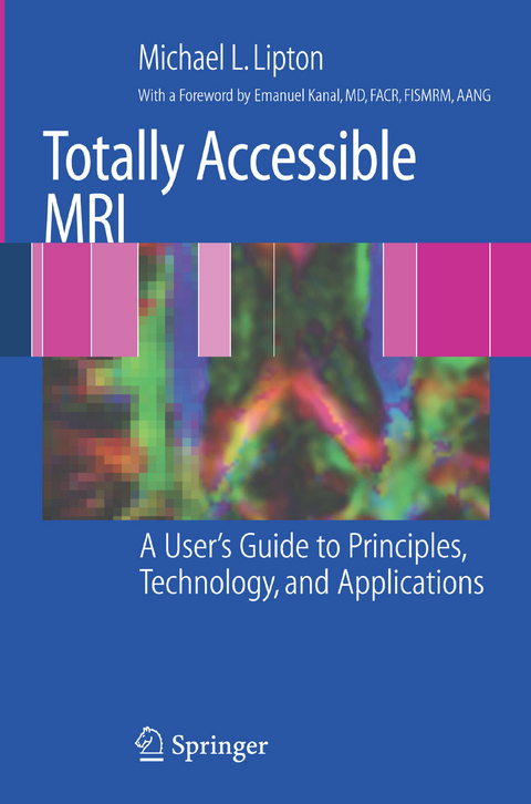
Totally Accessible MRI
Springer-Verlag New York Inc.
978-0-387-48895-0 (ISBN)
In the Beginning: Generating, Detecting, and Manipulating the MR (NMR) Signal.- Laying the Foundation: Nuclear Magnetism, Spin, and the NMR Phenomenon.- Rocking the Boat: Resonance, Excitation, and Relaxation.- Relaxation: What Happens Next?.- Image Contrast: T1, T2, T2*, and Proton Density.- Hardware, Especially Gradient Magnetic Fields.- User Friendly: Localizing and Optimizing the MRI Signal for Imaging.- Spatial Localization: Creating an Image.- Defining Image Size and Spatial Resolution.- Putting It All Together: An Introduction to Pulse Sequences.- Understanding, Assessing, and Maximizing Image Quality.- Artifacts: When Things Go Wrong, It’s Not Necessarily All Bad.- Safety: First, Do No Harm.- To the Limit: Advanced MRI Applications.- Preparatory Modules: Saturation Techniques.- Readout Modules: Fast Imaging.- Volumetric Imaging: The Three-dimensional Fourier Transform.- Parallel Imaging: Acceleration with SENSE and SMASH.- Flow and Angiography: Artifacts and Imaging of Coherent Motion.- Diffusion: Detection of Microscopic Motion.- Understanding and Exploiting Magnetic Susceptibility.- Spectroscopy and Spectroscopic Imaging: In Vivo Chemical Assays by Exploiting the Chemical Shift.
| Vorwort | E. Kanal |
|---|---|
| Zusatzinfo | 7 Illustrations, color; 146 Illustrations, black and white; XXII, 384 p. 153 illus., 7 illus. in color. |
| Verlagsort | New York, NY |
| Sprache | englisch |
| Maße | 155 x 235 mm |
| Themenwelt | Medizinische Fachgebiete ► Radiologie / Bildgebende Verfahren ► Kernspintomographie (MRT) |
| Medizinische Fachgebiete ► Radiologie / Bildgebende Verfahren ► Radiologie | |
| Naturwissenschaften ► Physik / Astronomie ► Angewandte Physik | |
| ISBN-10 | 0-387-48895-2 / 0387488952 |
| ISBN-13 | 978-0-387-48895-0 / 9780387488950 |
| Zustand | Neuware |
| Haben Sie eine Frage zum Produkt? |
aus dem Bereich


