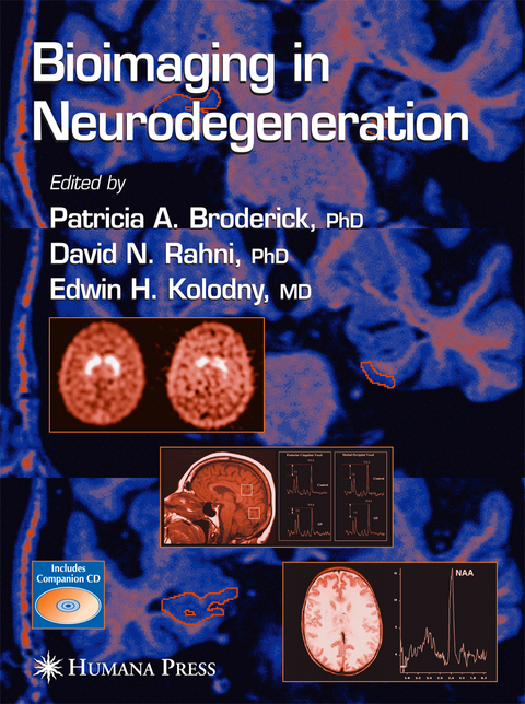
Bioimaging in Neurodegeneration
Humana Press Inc.
978-1-58829-391-6 (ISBN)
Parkinson’s Disease.- Magnetic Resonance Imaging and Magnetic Resonance Spectroscopy in Parkinson’s Disease.- Positron Emission Tomography and Single-Photon Emission Tomography in the Diagnosis of Parkinson’s Disease.- Positron Emission Tomography in Parkinson’s Disease.- [123I]-Altropane SPECT.- Positron Emission Tomography and Embryonic Dopamine Cell Transplantation in Parkinson’s Disease.- Alzheimer’s Disease.- Neurotoxicity of the Alzheimer’s ?-Amyloid Peptide.- Functional Imaging and Psychopathological Consequences of Inflammation in Alzheimer’s Dementia.- Neurotoxic Oxidative Metabolite of Serotonin.- Predicting Progression of Alzheimer’s Disease With Magnetic Resonance.- Stages of Brain Functional Failure in Alzheimer’s Disease.- Epilepsy.- Neocortical Epilepsy.- Pediatric Cortical Dysplasia.- Bioimaging L-Tryptophan in Human Hippocampus and Neocortex.- In Vivo Intrinsic Optical Signal Imaging of Neocortical Epilepsy.- Intraoperative Magnetic Resonance Imaging in the Surgical Treatment of Epilepsy.- Periodic Epileptiform Discharges Associated With Increased Cerebral Blood Flow.- Imaging White Matter Signals in Epilepsy Patients.- Leukodystrophy (White Matter) Diseases.- Overview of the Leukoencephalopathies.- Pyramidal Tract Involvement in Adult Krabbe’s Disease.- Imaging Leukodystrophies.- Advanced Magnetic Resonance Imaging in Leukodystrophies.- Childhood Mitochondrial Disorders and Other Inborn Errors of Metabolism Presenting With White Matter Disease.- Mitochondrial Disease.
| Erscheint lt. Verlag | 15.4.2005 |
|---|---|
| Reihe/Serie | Contemporary Neuroscience |
| Zusatzinfo | XVI, 313 p. With CD-ROM. |
| Verlagsort | Totowa, NJ |
| Sprache | englisch |
| Maße | 210 x 280 mm |
| Themenwelt | Medizin / Pharmazie ► Medizinische Fachgebiete ► Neurologie |
| Medizin / Pharmazie ► Studium | |
| Naturwissenschaften ► Biologie ► Humanbiologie | |
| ISBN-10 | 1-58829-391-2 / 1588293912 |
| ISBN-13 | 978-1-58829-391-6 / 9781588293916 |
| Zustand | Neuware |
| Haben Sie eine Frage zum Produkt? |
aus dem Bereich


