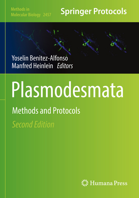
Plasmodesmata
Springer-Verlag New York Inc.
978-1-0716-2134-9 (ISBN)
Authoritative and up-to-date, Plasmodesmata: Methods and Protocols, Second Edition serves as a vital guide for all plant scientists, both novice and expert, especially those studying the intricacies of cell-to-cell communication pathways.
Plasmodesmata Structural Components and Their Role in Signaling and Plant Development.- Function of Plasmodesmata in the Interaction of Plants with Microbes and Viruses.- Plasmodesmata Ultrastructure Determination Using Electron Tomography.- Ultrastructural Analysis and Three-Dimensional Reconstruction of Plasmodesmata.- Serial Block Electron Microscopy to Study Plasmodesmata in the Vasculature of Arabidopsis thaliana Roots.- Focused Ion Beam-Scanning Electron Microscopy for Investigating Plasmodesmal Densities.- Measuring Plasmodesmata Density on Cell Interfaces of Monocot Leaves Using 3D Immunolocalization and Scanning Electron Microscopy.- Super-Resolution Imaging of Plasmodesmata Using 3D-Structured Illumination Microscopy.- In Vivo Aniline Blue Staining and Semi-Automated Quantification of Callose Deposition at Plasmodesmata.- Immunofluorescence Detection of Callose in Plant Tissue Sections.- Callose Detection and Quantification at Plasmodesmata in Bryophytes.-Isolation of Plasmodesmata Membranes for Lipidomic and Proteomic Analysis.- Methods for Detection of Protein Interactions with Plasmodesmata-Localized Reticulons.- Studying Protein-Protein Interactions at Plasmodesmata by Measuring Förster Resonance Energy Transfer.- Quantifying the Organization and Dynamics of the Plant Plasma-Membrane across Scales Using Light Microscopy.- Using Steady-State Fluorescence Anisotropy to Study Protein Clustering.- Quantification of Cell-to-Cell Connectivity Using Particle Bombardment.- Investigating Plasmodesmata Function in Arabidopsis thaliana Using a Low-Pressure Bombardment System and GFP Movement Assay.- Quantifying Plasmodesmatal Transport with an Improved GFP Movement Assay.- An Arabidopsis Callus Grafting Method to Test Cell-to-Cell Mobility of Proteins.- Quantification of Plasmodesmata Permeability in Arabidopsis Leaves by Tracing the Movement of GFP.- Tracking Intercellular Movement of Fluorescent Proteins in Bryophytes.- Virus Genome-Based Reporter for Analyzing Viral Movement Proteins and Plasmodesmata Permeability.- Analysis of the Distribution of Symplasmic Tracers during Zygotic and Somatic Embryogenesis.- Quantifying Intercellular Movement and Protein Stoichiometry for Computational Modeling.- Spatiotemporal Specific Blocking of Plasmodesmata by Callose Induction.- A Forward Genetic Approach to Identify Plasmodesmal Trafficking Regulators Based on Trichome Rescue.- In Vivo Visualization of Mobile mRNA Particles in Plants Using BglG.- Multi-Angle In Vivo Imaging of the Arabidopsis thaliana Shoot Apical Meristem (SAM).- More Insights from Ultrastructural and Functional Plasmodesmata Data Using PDinsight.- Measuring Intercellular Interface Area in Plant Tissues Using Quantitative 3D Image Analysis.
| Erscheinungsdatum | 04.04.2023 |
|---|---|
| Reihe/Serie | Methods in Molecular Biology |
| Zusatzinfo | 87 Illustrations, color; 3 Illustrations, black and white; XIII, 470 p. 90 illus., 87 illus. in color. |
| Verlagsort | New York, NY |
| Sprache | englisch |
| Maße | 178 x 254 mm |
| Themenwelt | Naturwissenschaften ► Biologie ► Botanik |
| Naturwissenschaften ► Biologie ► Zellbiologie | |
| Schlagworte | Callose accumulation • Nanochannels • Plant development and disease • Plasmodesmata architectures • Trafficking regulators • Vascular interface |
| ISBN-10 | 1-0716-2134-3 / 1071621343 |
| ISBN-13 | 978-1-0716-2134-9 / 9781071621349 |
| Zustand | Neuware |
| Informationen gemäß Produktsicherheitsverordnung (GPSR) | |
| Haben Sie eine Frage zum Produkt? |
aus dem Bereich


