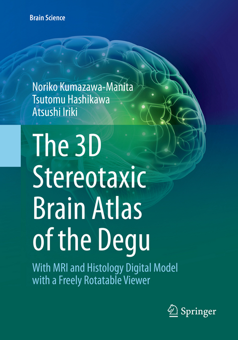
The 3D Stereotaxic Brain Atlas of the Degu
With MRI and Histology Digital Model with a Freely Rotatable Viewer
Seiten
2019
|
Softcover reprint of the original 1st ed. 2018
Springer Verlag, Japan
978-4-431-56866-7 (ISBN)
Springer Verlag, Japan
978-4-431-56866-7 (ISBN)
This book is the first digital atlas of the degu brain with microscopic features simultaneously in Nissl sections and magnetic resonance imaging (MRI). As an experimental animal model, the degu contributes to a variety of medical research fields in diabetes, hyperglycemia, pancreatic function, and adaptation to high altitude, among others. Recently the degu has gained increasing importance in the field of neuroscience, particularly in studies evaluating the relationship between sociality and cognitive brain functions, and in studies pertaining to the evolutional aspects of the acquisition of tool-use abilities. Furthermore, aging-related brain dysfunction in humans can be studied using this animal model in addition to mammals with much longer lifespans. This brain atlas is constructed to provide histological and volume-rendered information simultaneously, fitting with any spatial coordination in brain positioning. It can be a useful guide to degus as well as to other rodents for studies of brain structures conducted using MRI or other contemporary examination methods with volume-rendering functions.
Noriko Kumazawa-Manita Laboratory for Symbolic Cognitive Development, RIKEN Brain Science Institute, Japan Tsutomu Hashikawa Laboratory for Symbolic Cognitive Development, RIKEN Brain Science Institute, Japan Atsushi Iriki Laboratory for Symbolic Cognitive Development, RIKEN Brain Science Institute, Japan
Chapter 1: Introduction, Materials and Methods, and References.- Chapter 2: List of Structures.- Chapter 3: The Degu Brain Atlas.- Chapter 4: SG-eye Operation Manual.- Index of Structures and Abbreviations.
| Erscheint lt. Verlag | 14.2.2019 |
|---|---|
| Reihe/Serie | Brain Science |
| Zusatzinfo | 144 Illustrations, color; 54 Illustrations, black and white; IX, 144 p. 198 illus., 144 illus. in color. |
| Verlagsort | Tokyo |
| Sprache | englisch |
| Maße | 178 x 254 mm |
| Themenwelt | Studium ► 1. Studienabschnitt (Vorklinik) ► Physiologie |
| Naturwissenschaften ► Biologie ► Humanbiologie | |
| Naturwissenschaften ► Biologie ► Zoologie | |
| Schlagworte | Atlas • brain • Degu • Histology • MRI |
| ISBN-10 | 4-431-56866-2 / 4431568662 |
| ISBN-13 | 978-4-431-56866-7 / 9784431568667 |
| Zustand | Neuware |
| Haben Sie eine Frage zum Produkt? |
Mehr entdecken
aus dem Bereich
aus dem Bereich


