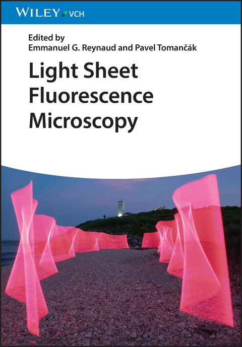
Light Sheet Fluorescence Microscopy
Wiley-VCH (Verlag)
978-3-527-34135-1 (ISBN)
An indispensable guide to a novel, revolutionary fluorescence microscopy technique!
Light sheet fluorescence microscopy is a revolutionary novel imaging technology, which drastically reduces phototoxicity and at the same time dramatically increases 3D resolution of the samples even when compared to confocal microscopy. These properties make the technology particularly suitable for imaging live samples. Since it is much less expensive but more powerful than a confocal microscope, light sheet fluorescence microscopy has become popular equipment in all laboratories which use fluorescent imaging of live samples, in particular cell biology, development biology and neurobiology labs.
This practical approach gives a comprehensive, hands-on overview of the basics of light sheet fluorescence microscopy, instrumentation, applications, sample preparation, and data analysis. The authors are the world leading experts in their corresponding fields and merge their expertises in physics, biology, and computer science in an unique book. It contains cutting edge methods and applications as well as valuable insider tips. A special feature of the book is discussion of a "do it yourself" light sheet microscope, making the technique affordable for each laboratory.
Emmanuel Reynaud has been Stokes Lecturer in Biology at University College Dublin, Ireland, since 2009. He received his Master's degree in molecular biology from the Victor Segalen University of Bordeaux, and his doctorate in biology from the University Paris XI-Orsay, France. In 2002, he received an EMBO Long Term Fellowship and moved to the European Molecular Biology Laboratory in Heidelberg, Germany, where he developed new methods in cell biology including laser nanosurgery approaches to study cytoskeleton dynamics as well as Golgi biogenesis. He was also involved in the development of the Light Sheet based Fluorescence Microscopy as a member of the Light Microscopy Group headed by Ernst H.K. Stelzer at EMBL. He has coordinated the Imaging Platform for the Tara Oceans Expedition and developed commercial applications with Carl Zeiss Microimaging, AURA Optik and ANDOR Ltd.
Pavel Tomancak is Senior Permanent Research Group Leader at the Max Planck Institute of Molecular Cell Biology and Genetics in Dresden, Germany. After studying Molecular Biology and Genetics at the Masaryk University in Brno, Czech Republic, he did his PhD at the European Molecular Biology Laboratory (EMBL) in the field of Drosophila developmental genetics. During his post-doctoral time at the University of California in Berkeley at the laboratory of Gerald M. Rubin, he established image-based genome-scale resources for patterns of gene expression in Drosophila embryos. His laboratory in Dresden continues to study patterns of gene expression during development by combining molecular, imaging and image analysis techniques. The group has lead a significant technological development aiming towards more complete quantitative description of gene expression patterns using light sheet microscopy.
LIGHT SHEET MICROSCOPY HARDWARE
Physics of Light Sheet-based Fluorescence Microscopes (e.g. Light Sheet formation, Optics, Detectors)
Overview of the Flavors of Light Sheet Microscopy (e.g. Arrangements of Optical Axes, Schematics, Commercial Set-ups, Extensions to Other Modalities)
Driving the Microscope and Open Access Electronics (e.g. Software to Pilot Light Sheet Microscopes (MicroManager), Arduino Solutions)
"Do It Yourself" Light Sheet based Microscope (e.g. OpenSPIM/OpenSPIN/LegoSPIM)
SAMPLE MOUNTING FOR LIGHT SHEET MICRSOCOPY
Sample Handling and Preparation (Including Sample Chamber Designs, Sample Positioning)
Clearing of Biological Tissues For Light Sheet Microscopy
Operation of Light Sheet-based Fluorescence Microscopes (Including In Multi-user Environment)
LIGHT SHEET MICRSOCOPY SOFTWARE
Image Processing of Multi-view Data (Registration, Fusion, and Deconvolution))
Visualization of Big Image Data (e.g. Local and Remote Viewing, 3D Rendering, Annotation)
Information Technology of Big Data Handling (e.g. Compression, Storage, Transfer)
Extracting Information From Light Sheet Datasets (e.g. Image Analysis Such As Atlas Registration, Segmentation, Tracking)
LIGHT SHEET MICROSCOPY APPLICATIONS
Applications In Developmental Biology
Applications In Cell Biology
Applications In Neurobiology
Applications In Biodiversity
LIGHT SHEET MICROSCOPY IMPLEMENTATIONS
Imaging Platform Integration
Laboratory Integration
IT Supports
| Erscheinungsdatum | 22.11.2018 |
|---|---|
| Verlagsort | Weinheim |
| Sprache | englisch |
| Maße | 170 x 244 mm |
| Gewicht | 788 g |
| Einbandart | kartoniert |
| Themenwelt | Medizin / Pharmazie ► Medizinische Fachgebiete ► Biomedizin |
| Medizin / Pharmazie ► Medizinische Fachgebiete ► Laboratoriumsmedizin | |
| Naturwissenschaften ► Biologie ► Biochemie | |
| Naturwissenschaften ► Biologie ► Genetik / Molekularbiologie | |
| Naturwissenschaften ► Chemie ► Analytische Chemie | |
| Schlagworte | Bild- u. Videoverarbeitung • Biowissenschaften • Cell & Molecular Biology • Chemie • Chemistry • Electrical & Electronics Engineering • Elektrotechnik u. Elektronik • Image and Video Processing • Life Sciences • Medical Science • Medizin • Microscopy • Mikroskopie • Radiologie • Radiologie u. Bildgebende Verfahren • Radiology & Imaging • Zell- u. Molekularbiologie |
| ISBN-10 | 3-527-34135-8 / 3527341358 |
| ISBN-13 | 978-3-527-34135-1 / 9783527341351 |
| Zustand | Neuware |
| Informationen gemäß Produktsicherheitsverordnung (GPSR) | |
| Haben Sie eine Frage zum Produkt? |
aus dem Bereich


