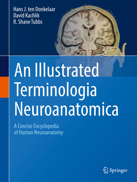
An Illustrated Terminologia Neuroanatomica
Springer International Publishing (Verlag)
978-3-319-64788-3 (ISBN)
This book is unique in that it provides the reader with the most up-to-date terminology used to describe the human nervous system (central and peripheral) and the related sensory organs, i.e., the Terminologia Neuroanatomica (TNA), the official terminology of the IFAA (International Federation of Associations of Anatomists). The book provides a succinct but detailed review of the neuroanatomical structures of the human body and will greatly benefit not only various specialists such as (neuro)anatomists, neurologists and neuroscientists, but also students taking neuroanatomy and neuroscience courses.
The book offers a high yield, combined presentation of neuroanatomical illustrations and text and provides the reader a 'one-stop source' for studying the intricacies of the human nervous system and its sensory organs. It includes an alphabetical list of official English terms and synonyms with the official Latin terms and synonyms from the TNA. With regard to the entries, the nameof the item in standardized English is provided, followed by synonyms and the official TNA Latin term, Latin synonyms and eponyms, a short description and in many cases one or more illustrations. To facilitate the use of illustrations, certain entries such as the gyri or sulci of the cerebral cortex are presented together with extensive cross-references. Terms that form part of a certain structure (such as the amygdaloid body, the thalamus and the hypothalamus) are listed under the respective structure. Segments and branches of arteries are discussed under the main artery, for example the A1-A5 segments under the anterior cerebral artery. Most nerves can be found following their origin from the brachial, cervical and lumbosacral plexuses. However, the major nerves of the limbs are discussed separately, as are the cranial nerves. Nuclei can be found by their English name or under Nuclei by their eponym.
Authors:Hans J. ten Donkelaar, M.D., Ph.D., 935 Department of Neurology, Donders Center forMedical Neuroscience, Radboud University Nijmegen Medical Center, has published three major works, not two; you probably forgot 'The Central NervousSystem of Vertebrates' (Nieuwenhuys, ten Donkelaar, Nicholson, Springer 1998) Prof. Kachlík is Head of the Department of Anatomy of the Second Faculty ofMedicine of Charles University in Prague. He is an expert on anatomical nomenclature. Prof. Tubbs serves as Chief Scientific Officer for the Seattle Foundation. He has published alarge number of papers, authored over 20 books, and is editor-in-chief of the journal Clinical Anatomy. Hans J. ten Donkelaar (1946) studied Medicine at the University of Nijmegen (TheNetherlands), where he received his M.D. (1974) and Ph.D. (1975). In 1978, he was appointedAssociate Professor of Neuroanatomy at the Department of Anatomy and Embryology of thatUniversity. His research interests are developmental and comparative aspects of motorsystems, developmental disorders of the CNS and neurodegenerative disease. With RudolfNieuwenhuys and Charles Nicholson he published The Central Nervous System of Vertebrates(1998, Springer) and with Anthony Lohman an anatomy and embryology textbook in Dutch,which is now in its fourth edition (ten Donkelaar HJ, Oostra R-J 2014 Klinische Anatomie enEmbryologie. Springer Media/Houten/NL). In 1998, he came to the Department of Neurologyof the Radboud University Nijmegen Medical Center to do research on developmental andneurodegenerative diseases. In 2006, he published with Martin Lammens and Akira HoriClinical Neuroembryology: Development and developmental disorders of the human centralnervous system (Springer), which is in its second edition now (2014), and in 2011 ClinicalNeuroanatomy: Brain circuitry and its disorders (Springer). Since 2012, he is Co-ordinator ofthe FIPAT (Federative International Programme for Anatomical Terminology) WorkingGroup Neuroanatomy and responsible for the TNA.David Kachlík (1974) studied Medicine in Prague, where he received his M.D. (1998) andPh.D. (2006). Since 1998, he worked in the Department of Anatomy of the Third Faculty ofMedicine of the Charles University in Prague. In 2016, he was appointed Professor ofAnatomy at its Second Faculty of Medicine. His research concerns vascular andmusculoskeletal anatomy, peripheral nerves and morphological terminology andnomenclature. In 2010, he published with Pavel Čech, Vladimír Musil and Václav Báča ČeskéTĕlovĕdné Názvosloví (Brno), the Czech translation of the Terminologia Anatomica of 1998.He is a member of FIPAT and published a series of influential papers on anatomicalterminology.R. Shane Tubbs (1969) received his MS (1998) and Ph.D (2002) at the University ofAlabama, Birmingham, AL, where he started his career as a clinical anatomist and researcher.His research focus has been on the so-called 'reverse translational anatomy research', whereclinical problems are identified and solved with anatomical studies. He has published a largenumber of papers, authored over 20 books, and is editor-in-chief of the journal ClinicalAnatomy. He has recently moved to Seattle, WA, as the Chief Scientific Officer at the SeattleScience Foundation, where he continues his research and teaching to medical professionals.He is an editor of the 41th edition of Gray's Anatomy and recently published withMohammadali M. Shoja and Marios Loukas Bergman's Comprehensive Encyclopedia ofHuman Anatomic Variation (Wiley, Blackwell, Hoboken, New Jersey, 2016). He is also a member of FIPAT. Contributors:Robert H. Baud, Ph.D., Service of Medical Informatics, University Hospitals of Geneva, CH-1211 Geneva 14, Switzerland (Informatics and cross-referencing) Axel Brehmer, M.D., Ph.D. Institute of Anatomy. University of Erlangen-Nuremberg, Germany, Krankenhausstraβe 9, D-91054 Erlangen, Germany (Enteric nervous system) Jonas Broman, Ph.D., Institute for Clinical and Experimental Medicine, University of Linköping, S-58183 Linköping, Sweden (Spinal cord and Ascending systems)Jean Büttner-Ennever, Ph.D., Department of Neuroanatomy, Ludwig-Maximilian-University Faculty of Medicine, Pettenkoferstrasse 11, D-80336 Munich, Germany (Brain stem nuclei)Matthew Carlson, M.D., Department of Otolaryngology, Mayo Clinic, 200 1st St SW, 55905 Rochester, MN, USA (Ear)Marco Catani, M.D., Department of Neuroimaging, King's College London, Strand, London WC2R2LS, UK (Association pathways, DTI)Andras Csillag, M.D., Ph.D., Department of Anatomy, Histology and Embryology, Semmelweis University, Tuzoltó utca 58, HU-1094 Budapest, Hungary (Basal ganglia)Anja K.E. Horn-Bochtler, Ph.D., Department of Anatomy and Cell Biology I, Ludwig-Maximilian-University Faculty of Medicine, Pettenkoferstrasse 11, D-80336 Munich, Germany (Brain stem nuclei)Ricardo Insausti, M.D., Ph.D., Department of Anatomy, Faculty of Medicine, Universidad Castilla-La Mancha, Avenida Almansa 14, E-02006 Albacete, Spain (Hippocampal formation and related structures)Geoffrey Meyer, Ph.D., School of Anatomy, Physiology and Human Biology, University of Western Australia, Crawley WA 6009, Australia (Histology)Veronika Němcová, M.D., Ph.D., Department of Anatomy, First Faculty of Medicine, Charles University, U nemocnice 3, 12800 Praha 2, Czech Republic (Gross anatomy and Brain stem)Luis Puelles, M.D., Ph.D., Department of Human Anatomy and Psychobiology, Faculty of Medicine, University of Murcia, E-30100 Murcia, Spain (Developmental aspects)Clifford B. Saper, M.D., Ph.D., Harvard University Press and Department of Neurology, Beth Israel Deaconess Medical Center, 330 Brookline Avenue, Boston, MA 02215, USA (Hypothalamus and Preoptic region)Gulgun Sengul, M.D., Ph.D., Department of Anatomy, Ege University School of Medicine, Bornova, 35100 Izmir, Turkey (Spinal cord)
| Erscheinungsdatum | 19.05.2018 |
|---|---|
| Zusatzinfo | XVII, 491 p. 583 illus., 351 illus. in color. |
| Verlagsort | Cham |
| Sprache | englisch |
| Maße | 210 x 279 mm |
| Gewicht | 1479 g |
| Themenwelt | Medizin / Pharmazie ► Medizinische Fachgebiete ► Neurologie |
| Studium ► 1. Studienabschnitt (Vorklinik) ► Anatomie / Neuroanatomie | |
| Naturwissenschaften ► Biologie ► Humanbiologie | |
| Schlagworte | anatomy • Biomedical and Life Sciences • Human nervous system • Medical Research • Neuroanatomy • neuroembryology • Neurohistology • Neurology • Neurology & clinical neurophysiology • Neurology & clinical neurophysiology • Neurosciences • sensory organs • Terminology |
| ISBN-10 | 3-319-64788-1 / 3319647881 |
| ISBN-13 | 978-3-319-64788-3 / 9783319647883 |
| Zustand | Neuware |
| Informationen gemäß Produktsicherheitsverordnung (GPSR) | |
| Haben Sie eine Frage zum Produkt? |
aus dem Bereich


