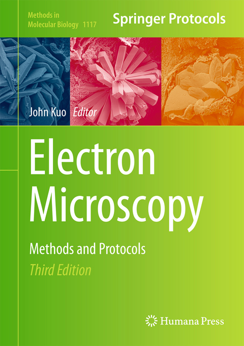
Electron Microscopy
Humana Press Inc. (Verlag)
978-1-62703-775-4 (ISBN)
Authoritative and practical, Electron Microscopy: Methods and Protocols, Third Edition provides the most up-to-date and essential information in electron microscopy techniques and methods provided in this edition will assist in advancing future molecular and biological research.
Conventional Specimen Preparation Techniques For Transmission Electron Microscopy of Cultured Cells.- Microwave-assisted Processing and Embedding for Transmission Electron Microscopy.- Processing Plant Tissues for Ultrastructural Study.- Staining Sectioned Biological Specimens for Transmission Electron Microscopy: Conventional and En Bloc Stains.- Metal Shadowing for Electron Microscopy.- Freeze Fracture and Freeze Etching.- Conventional Specimen Preparation Techniques For Scanning Electron Microscopy of Biological Specimens.- High-pressure Freezing: Current State and Future Prospects.- Cryo-fixation by Self-pressurized Rapid Freezing.- Cryo-Electron Microscopy of Vitreous Sections.- Negative Staining and Cryo-negative Staining: Applications in Biology and Medicine.- Electron Microscopy of the Microtubule Cytoskeleton Assembly Vitro.- Cryosectioning Fixed and Cryoprotected Biological Material for Immmunocytochemistry.- Analysis of Specificity in Immunoelectron Microscopy.- Cryo-Electron Microscopy of Membrane Proteins.- Tracking DNA and RNA Sequences at High Resolution.- Visualization of DNA and Protein-DNA Complexes with Atomic Force Microscopy.- Biological Applications of Phase-Contrast Electron Microscopy.- Single Particle Cryo-Electron Microscopy And 3-D Reconstruction Of Viruses.- Electron Tomography for Organelles, Cells and Tissues.- Correlative Light and Electron Microscopy: from Live Cell Dynamic to 3D Ultrastructure.- Nanometer-resolution Fluorescence Electron Microscopy (Nano-EM) in Cultured Cells.- Correlative Fluorescence- and Electron Microscopy of Quantum Dot Labeled Proteins on Whole Cells in Liquid.- FIB-SEM Tomography in Biology.- Correlative Light and Electron Microscopy using Immunolabeled Sections.- Correlative 3D Imaging: CLSM and FIB-SEM Tomography Using High-Pressure Frozen, Freeze-Substitued Biological Samples.- Three-dimensional Imaging of Adherent Cells using FIB-SEM and STEM.- X-Ray Microanalysis in the Scanning Electron Microscope.- Application of SEM and EDX in Studying Biomineralization in Plant Tissues.- Freeze Stabilization and Cryopreparation Technique for Visualizing the Water Distribution in Woody Tissues by X-ray Imaging and Cryoscanning Electron Microscopy.- Biological Applications of Energy-Filtered TEM.- Secondary Ion Mass Spectrometry Imaging of Biological Cells and Tissues.- Elemental and Isotopic Imaging of Biological Samples using Nano SIMS.- 3D Chemical Mapping: Application of Scanning Transmission (soft) X-ray Microscopy (STXM) in Combination with Angle-Scan Tomography in Bio-, Geo- and Environmental Science.
| Reihe/Serie | Methods in Molecular Biology ; 1117 |
|---|---|
| Zusatzinfo | 100 Illustrations, color; 156 Illustrations, black and white; XVIII, 799 p. 256 illus., 100 illus. in color. |
| Verlagsort | Totowa, NJ |
| Sprache | englisch |
| Maße | 178 x 254 mm |
| Themenwelt | Medizinische Fachgebiete ► Radiologie / Bildgebende Verfahren ► Radiologie |
| Naturwissenschaften ► Biologie ► Genetik / Molekularbiologie | |
| Schlagworte | bio-molecular electron microscopy • bio-molecular research • correlative microscopy • Cryo-specimen • cryoTEM 3D tomography • DNA and RNA tracking • electron microscopes • (EM) data • Focussed Ion Beam (FIB) • scanning electron microscope (SEM) • Transmission Electron Microscope (TEM) tomography |
| ISBN-10 | 1-62703-775-6 / 1627037756 |
| ISBN-13 | 978-1-62703-775-4 / 9781627037754 |
| Zustand | Neuware |
| Haben Sie eine Frage zum Produkt? |
aus dem Bereich


