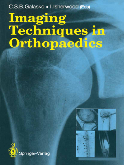
Imaging Techniques in Orthopaedics
Springer London Ltd (Verlag)
978-1-4471-1642-4 (ISBN)
Recent years have witnessed major developments in diagnostic imaging methods. The facilities for these new methods are sometimes expensive. and not always accessible. yet they continue to improve and to change. It is essential that those concerned with orthopaedic imaging should appreciate not only recent developments but also the changes likely to occur during the next few years. It is also important that the indications. contraindications. uses and complications for each individual imaging technique should be understood. This book is an attempt to provide such information for orthopaedic surgeons. diagnostic radiologists. and other clinicians. par ticularly those in training or those who are involved in management of patients with disorders of the musculoskeletal system. In the first part of the book the different imaging techniques are discussed. with emphasis on advantages and disadvantages. indications and contraindica tions. In the second part. authors have been asked to discuss ways in which specific groups of disorders might be investigated. It is hoped that the reader will obtain from this section a balanced view of the different diagnostic imaging methods. the indications for their use. and the sequence in which they might be carried out. The Editors are grateful to aU authors for the time and work they have put into their individual chapters. They are also grateful to the publishers. in particular Michael Jackson. for help given in the preparation of this book. Manchester C. S. B. Galasko I.
1 Radiological Techniques.- 1 Conventional Radiography of the Appendicular Skeleton.- 2 Angiography of the Appendicular Skeleton.- 3 Arthrography.- 4 Conventional Radiography of the Axial Skeleton.- 5 Myelography.- 6 Epidurography.- 7 Angiography of the Axial Skeleton.- 8 Discography.- 9 Facet Arthrography.- 10 Computed Tomography.- 11 Digital Orthopaedic Radiography: Vascular and Non-vascular.- 12 Magnetic Resonance Imaging (MRI).- 13 Skeletal Scintigraphy.- 14 Isotope Techniques in the Investigation of Diseases of Joints.- 15 Ultrasound of the Axial Skeleton.- 16 Appendicular Ultrasound.- 2 Bone Mineral Studies.- 17 Bone Measurement by Conventional Radiographic Techniques.- 18 Photon Absorptiometry.- 19 Quantitative Computed Tomography (QCT).- 3 Clinical Indications.- 20 Musculoskeletal Trauma.- 21 Primary Tumours of Bone and Soft Tissue.- 22 Metastatic Tumours.- 23 Infection.- 24 Skeletal Dysplasias.- 25 Back Pain.- 26 Spinal Deformity.- 27 Disorders in Childhood.- 28 Osteonecrosis.
| Zusatzinfo | XI, 376 p. |
|---|---|
| Verlagsort | England |
| Sprache | englisch |
| Maße | 210 x 280 mm |
| Themenwelt | Medizinische Fachgebiete ► Chirurgie ► Unfallchirurgie / Orthopädie |
| Medizinische Fachgebiete ► Radiologie / Bildgebende Verfahren ► Radiologie | |
| Medizin / Pharmazie ► Studium ► 1. Studienabschnitt (Vorklinik) | |
| Naturwissenschaften ► Biologie ► Biochemie | |
| ISBN-10 | 1-4471-1642-9 / 1447116429 |
| ISBN-13 | 978-1-4471-1642-4 / 9781447116424 |
| Zustand | Neuware |
| Informationen gemäß Produktsicherheitsverordnung (GPSR) | |
| Haben Sie eine Frage zum Produkt? |
aus dem Bereich


