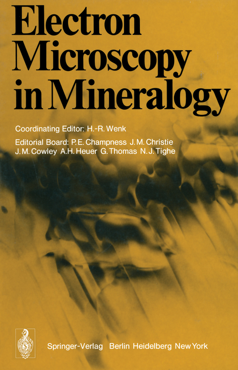
Electron Microscopy in Mineralogy
Springer Berlin (Verlag)
978-3-642-66198-3 (ISBN)
Section 1 Introduction.- Section 2 Contrast.- 2.1 Fundamentals of Electron Microscopy.- 2.2 Interpretation of Electron Diffraction Patterns.- 2.3 Contrast Effects at Planar Interfaces.- 2.4 Computer Simulation of Dislocation Images in Quartz.- 2.5 The Direct Imaging of Crystal Structures.- 2.6 A Comparison of Bright Field and Dark Field Imaging of Pyrrhotite Structures.- Section 3 Experimental Techniques.- Section 4 Exsolution.- 4.1 Exsolution in Silicates.- 4.2 Coarsening in a Spinodally Decomposing System: TiO2-SnO2.- 4.3 Magnetite Lamellae in Reduced Hematites.- 4.4 Precipitation in the Ilmenite-hematite System.- 4.5 Pigeonite Exsolution from Augite.- 4.6 The Transformation of Pigeonite to Orthopyroxene.- 4.7 On the Detailed Structure of Ledges in an Augite-enstatite Interface.- 4.8 The Phase Distributions in Some Exsolved Amphiboles.- 4.9 Physical Aspects of Exsolution in Natural Alkali Feldspars.- 4.10 Analytical Electron Microscopy of Exsolution Lamellae in Plagioclase Feldspars.- 4.11 Exsolution in Metamorphic Bytownite.- Section 5 Polymorphic Phase Transitions.- 5.1 Polymorphic Phase Transitions in Minerals.- 5.2 Direct Observation of Iron Vacancies in Polytypes of Pyrrhotite.- 5.3 Rutile: Planar Defects and Derived Structures.- 5.4 High-resolution Electron Microscopy of Unit Cell Twinning in Enstatite.- 5.5 Polytypism in Wollastonite.- 5.6 High-resolution Electron Microscopy of Labradorite Feldspar.- 5.7 Origin of the (c) Domains of Anorthite.- 5.8 On Polymorphism of BaAl2Si2O8.- 5.9 The Submicroscopic Structure of Wenkite.- Section 6 Deformation Defects.- 6.1 Deformation Structures in Minerals.- 6.2 Work Hardening and Creep Deformation of Corundum Single Crystals.- 6.3 Dislocation Structures in Synthetic Quartz.- 6.4 The Microstructure of Some Naturally Deformed Quartzites.- 6.5 Defects in Deformed Calcite and Carbonate Rocks.- 6.6 Plasticity of Olivine in Peridotites.- 6.7 The Role of Crystal Defects in the Shear-induced Transformation of Orthoenstatite to Clinoenstatite.- Section 7 Special Techniques and Applications.- 7.1 Amorphous Materials.- 7.2 Signals Excited by the Scanning Beam.- 7.3 Analytical Electron Microscopy of Minerals.- 7.4 X-ray Microanalysis Using a Scanning Electron Microscope.- 7.5 Quantitative X-ray Microanalysis of Thin Foils.- 7.6 Particle Track Studies.- 7.7 Stony Meteorites.- 7.8 Microcracks in Crystalline Rocks.
| Erscheint lt. Verlag | 18.11.2011 |
|---|---|
| Mitarbeit |
Stellvertretende Herausgeber: P.E. Champness, J.M. Christie, J.M. Cowley, A.H. Heuer, G. Thomas, N.J. Tighe Chef-Herausgeber: H.-R. Wenk |
| Zusatzinfo | XIV, 566 p. |
| Verlagsort | Berlin |
| Sprache | englisch |
| Maße | 170 x 244 mm |
| Gewicht | 993 g |
| Themenwelt | Naturwissenschaften ► Chemie |
| Naturwissenschaften ► Geowissenschaften ► Geologie | |
| Naturwissenschaften ► Geowissenschaften ► Mineralogie / Paläontologie | |
| Schlagworte | Crystal • crystallography • Electron Microscope • electron microscopy • Elektronenmikroskopie • Mineralogie • Mineralogy • Transmission Electron Microscopy |
| ISBN-10 | 3-642-66198-X / 364266198X |
| ISBN-13 | 978-3-642-66198-3 / 9783642661983 |
| Zustand | Neuware |
| Haben Sie eine Frage zum Produkt? |
aus dem Bereich


