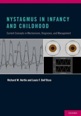
Nystagmus In Infancy and Childhood
Oxford University Press Inc (Verlag)
978-0-19-985700-5 (ISBN)
Nystagmus in Infancy and Childhood is a highly-illustrative and thoughtfully written text that provides clinicians and scientists with detailed yet concise information regarding our current understanding, evaluation, and treatments of nystagmus in infancy and childhood. Throughout the text are clinical pearls and narrative observations intended to help the reader appreciate the enormous strides forward in the past 50 years of nystagmus research.
Timely and comprehensive, this book is an "everything you need to know" resource, and will provide the reader with:
- detailed methodologies of investigation, including analysis software, models of the ocular motor system, and current hypotheses regarding ocular motor oscillations
- complementary appendices that can be used for special purposes, i.e., as clinical examination sheets, patient information sheets, and algorithm for computer analysis of nystagmus waveforms
- new therapeutic approaches, using relevant eye-movement data and mechanisms
- a roadmap toward a more rational, data-driven approach to the medical management of infantile nystagmus
As the only resource effectively comprising the past 50 years of nystagmus research and therapeutic implications, Nystagmus in Infancy and Childhood will be a comprehensive and invaluable guide to for both clinicians and scientists who care for infants and children with nystagmus.
Richard W. Hertle MD, FACS, FAAO, FAAP, is Chief of Pediatric Ophthalmology and Director of Adult Motility at The Laboratory of Visual and Ocular Motor Physiology, Children's Hospital Medical Center of Akron, as well as Professor of Ophthalmology at SUMMA Health System, Akron, Ohio and The Northeastern Ohio Universities College of Medicine, Rootstown, OH. Louis F. Dell'Osso, PhD, is Professor of Neurology and Biomedical Engineering at Case Western Reserve University and Laboratory, Louis Stokes Cleveland, VA Medical Center.
Chapter 1. Relevant Anatomy and Physiology ; 1.1 INFRANUCLEAR OCULAR MOTOR ANATOMY ; 1.1.1 Extraocular Muscles ; 1.1.2 Extraocular Muscle Pulleys ; 1.1.3 Orbital Tissues ; 1.2 SUPRANUCLEAR OCULAR MOTOR ANATOMY ; 1.2.1 Frontal Eye Fields ; 1.2.2 Superior Colliculus ; 1.2.3 Brainstem Nuclei ; 1.2.4 Vestibular Nuclei ; 1.2.5 Cerebellum ; 1.3 AFFERENT SYSTEM ; 1.3.1 Retina/Optic Nerve ; 1.3.2 Optic Nerve ; 1.3.3 Lateral Geniculate ; 1.3.4 Geniculostriate ; 1.3.5 Association Cortex ; 1.3.6 Ocular Motor Proprioception ; 1.4 EFFERENT SYSTEM ; 1.4.1 Smooth Pursuit System ; 1.4.2 Saccadic System ; 1.4.3 Vergence System ; 1.4.4 Vestibuloocular System ; Chapter 2. Infantile Nystagmus SyInfantile Nystagmus Syndromendrome ; 2.1 CHARACTERISTICS OF INS ; 2.1.1 History and Background ; 2.1.1.1 Ancient Descriptions and Theories ; 2.1.1.2 Connection to Fixation Attempt ; 2.1.1.3 Modern Physiological Investigation ; 2.1.2 Waveforms, Models, and Mechanisms ; 2.1.2.1 Waveform Types ; 2.1.2.2 Braking and Foveating Saccades ; 2.1.2.3 The Foveation Period ; 2.1.2.4 Foveation Accuracy ; 2.1.2.5 Target Acquisition Time ; 2.1.2.6 Smooth Pursuit ; 2.1.3 The Static Neutral Zone/Region ; 2.1.3.1 Latent Component ; 2.1.4 The Dynamic Neutral Zone/Region ; 2.1.4.1 Asymmetric, (a)Periodic Alternation ; 2.1.4.2 Optokinetic, Pursuit and Vestibuloocular Responses ; 2.1.5 The Null Angle/Zone/Region ; 2.1.6 The Convergence Null ; 2.1.7 The Saccadic Response ; 2.1.8 Static and Dynamic Head Posturing ; 2.1.9 Foveation and Visual Acuity (High Spatial Frequency Vision) ; 2.1.9.1. The eXpanded Nystagmus Acuity Function (NAFX) ; 2.1.9.1.1 NAFX vs. Gaze Angle ; 2.1.9.1.2 LFD and TID ; 2.1.10 Oscillopsia Suppression ; 2.1.10.1 Foveation Dynamics ; 2.1.10.2 Temporal Sampling ; 2.1.10.3 Efference Copy ; 2.1.11 Afferent Stimulation ; 2.1.11.1 Cutaneous Trigeminal Stimulation ; 2.1.11.2 Deep Muscle Stimulation ; 2.1.11.3 Contact Lenses ; 2.1.11.4 Biofeedback ; 2.1.12 Canine Nystagmus (Achiasmatic Belgian Sheepdog) ; 2.1.12.1 See-Saw ; 2.1.12.2 Pendular ; 2.1.12.3 Tenotomy & Reattachment Procedure ; 2.1.13 Canine Model of INS with RPE65 Retinal Degeneration (Briard) ; 2.2 ETIOLOGY OF INS ; 2.2.1 Familial (Gene Defect) ; 2.2.2 Developmental Disturbance Of Ocular Motor System with Associated Sensory System Deficit. ; 2.2.2.1 Albinism ; 2.2.2.2 Achiasma ; 2.2.2.3 Infantile Strabismus ; 2.2.2.4 Non-vectorial Visual Sensory Deficits ; 2.2.3 The Direct Cause(s) of INS with or without Sensory/Genetic Deficits ; 2.2.3.1 Loss of Smooth-Pursuit Damping ; 2.2.3.2 Tonic Vestibular-Optokinetic Imbalance ; 2.3 VISUAL FUNCTION DEFICITS AND MEASUREMENTS OF INS ; 2.3.1 Static Deficits ; 2.3.1.1 The eXpanded Nystagmus Acuity Function and Longest Foveation ; Domain Measures ; 2.3.2 Dynamic Deficits ; 2.3.2.1 Target Acquisition Time (Stationary Targets) ; 2.3.2.2 Target Acquisition Time (Moving Targets) ; 2.3.3 Clinical ; 2.3.3.1 Visual Acuity at Different Gaze Angles ; 2.4 TREATMENTS OF INS ; 2.4.1 Goals ; 2.4.2 Non-Surgical ; 2.4.2.1 Prisms ; 2.4.2.2 Contact Lenses ; 2.4.2.3 Drugs ; 2.4.2.4 Biofeedback ; 2.4.2.5 Gene-Transfer Therapy ; 2.4.3 Surgical ; 2.4.3.1 Four-Muscle Resection & Recession Procedure (Operation 1) ; (aka Kestenbaum, Anderson-Kestenbaum, or Anderson+Goto) ; 2.4.3.2 Two-Muscle Recession Procedure (Operation 1A) (aka Anderson) ; 2.4.3.3 Bilateral Medial Rectus Recession Procedure (Operation 8) ; 2.4.3.4 Tenotomy & Reattachment Procedure (Operation 6) ; Chapter 3. Fusion Maldevelopment Nystagmus Syndrome ; 3.1 CHARACTERISTICS OF FMNS ; 3.1.1 Waveforms, Models, and Mechanisms ; 3.1.1.1 Types (FMNS plus Nucleus of the Optic Tract) ; 3.1.1.2 The Fixating Eye ; 3.1.1.3 Target Foveation and Dual-Mode Fast Phases ; 3.1.1.4 Foveation Accuracy ; 3.1.2 Variation with Gaze Angle ; 3.1.3 Head Position ; 3.1.4 Foveation, NAFX, and Acuity ; 3.1.5 Efference Copy, Foveation, and Oscillopsia Suppression ; 3.2 TREATMENTS OF FMNS ; 3.2.1. Fixation Preference ; 3.2.2. Alexander's Law ; 3.2.3. Eye Muscle Surgery ; Chapter 4. Other Types of Nystagmus of Infancy ; 4.1 NYSTAGMUS BLOCKAGE SYNDROME ; 4.1.1 Characteristics of NBS ; 4.1.1.1 Multiple Types of Nystagmus ; 4.1.1.2 Waveforms and Mechanisms ; 4.1.1.2.1 Target Foveation ; 4.1.1.2.2 Foveation Accuracy ; 4.1.1.3 Purposive Esotropia ; 4.1.1.4 Head Position ; 4.1.1.5 Blockage Syndrome Types I and II ; 4.1.1.6 Foveation, NAFX, and Acuity ; 4.1.1.7 Efference Copy, Foveation, and Oscillopsia Suppression ; 4.1.2 Treatments of NBS ; 4.1.2.1. Fixation Preference ; 4.1.2.2. Alexander's Law ; 4.1.2.3. Surgical ; 4.1.2.3.1. Fixating Eye ; 4.2 SPASMUS NUTANS SYNDROME ; 4.2.1 Characteristics of SNS ; 4.2.1.1 Waveforms and Mechanisms ; 4.2.1.2 Variable Interocular Phase ; 4.2.1.3 Head Nodding ; 4.2.2 Treatment of SNS ; Chapter 5. Differential Diagnosis of Nystagmus In Infancy and Childhood ; 5.1 NYSTAGMUS WITHOUT ASSOCIATED NEUROLOGICAL DISEASE-<"BENIGN>" ; 5.1.1. Infantile Nystagmus Syndrome ; 5.1.1.1 Association with Strabismus ; 5.1.1.2 Clinical Signs and Symptoms ; 5.1.1.3 Differential Diagnosis ; 5.1.1.4 Reverse-Cover and Gaze-Angle Cover Tests ; 5.1.2 Fusion Maldevelopment Nystagmus Syndrome ; 5.1.2.1 Association with Strabismus ; 5.1.2.2 Clinical Signs and Symptoms ; 5.1.2.3 Differential Diagnosis ; 5.1.2.4 Alternate-Cover and Gaze-Angle Cover Tests ; 5.1.3 Nystagmus Blockage Syndrome ; 5.1.3.1 Association with Strabismus ; 5.1.3.2 Clinical Signs and Symptoms ; 5.1.3.3 Differential Diagnosis ; 5.1.4 Spasmus Nutans Syndrome ; 5.1.4.1 Association with Strabismus ; 5.1.4.2 Clinical Signs and Symptoms ; 5.1.4.3 Differential Diagnosis ; 5.1.5 Nystagmus and Strabismus ; 5.2 NYSTAGMUS WITH ASSOCIATED NEUROLOGICAL DISEASE-<"SYMPTOMATIC>" ; 5.2.1 Vestibular Nystagmus ; 5.2.1.1 Peripheral Vestibular Imbalance ; 5.2.1.2 Central Vestibular Imbalance ; 5.2.1.3 Central Vestibular Instability (Periodic Alternating) ; 5.2.2 Gaze-holding Deficiency Nystagmus ; 5.2.2.1 Eccentric Gaze, Gaze-evoked, Rebound ; 5.2.2.2 Gaze Instability (<"Run-away>") ; 5.2.3 <"Vision-Loss>" Nystagmus ; 5.2.3.1 Pre-chiasmal, Optic Chiasm, Post-chiasmal Vision Loss ; 5.2.4 Other Pendular Nystagmus Associated with Diseases of Central Myelin ; 5.2.4.1 Oculopalatal Tremor or <"Myoclonus>" ; 5.2.4.2 Pendular Vergence Nystagmus Associated with Whipple's Disease ; 5.2.5 Convergence/Convergence Evoked Nystagmus ; 5.2.6 Upbeat Nystagmus ; 5.2.7 Downbeat Nystagmus ; 5.2.8 Torsional Nystagmus ; 5.2.9 <"See-Saw>" Nystagmus ; 5.2.10 Lid Nystagmus ; 5.3 SACCADIC INTRUSIONS/OSCILLATIONS ; 5.3.1 Square Wave Jerks And Oscillations ; 5.3.2 Square-Wave Pulses ; 5.3.3 Staircase Saccadic Intrusions ; 5.3.4 Macrosaccadic Oscillations ; 5.3.5 Saccadic Pulses (Single And Double) ; 5.3.6 Convergence Retraction <"Nystagmus>" ; 5.3.7 Dissociated Ocular Oscillations ; 5.3.8 Dysmetric Saccades ; 5.3.9 Ocular Flutter ; 5.3.10 Flutter Dysmetria ; 5.3.11 Opsoclonus ; 5.3.11.1 Opsoclonus-Myoclonus ; 5.3.12 Superior Oblique Myokymia ; 5.3.13 Ocular bobbing ; 5.3.13.1 Typical ; 5.3.13.2 Monocular ; 5.3.13.3 Atypical ; 5.3.14 Psychogenic (Voluntary) Flutter ; Chapter 6. Afferent Visual System - Clinical Examination Procedures ; 6.1 SUBJECTIVE TESTING ; 6.1.1 Teller Acuity Card Procedure ; 6.1.2 Visual Acuity Testing (High Spatial Frequency Vision) ; 6.1.3 Stereo Testing ; 6.1.4 Color-Vision Testing ; 6.1.5 Contrast-Sensitivity Testing ; 6.1.6 Gaze- and Time-Dependent Acuity Testing ; 6.1.7 Visual Field Testing ; 6.2 OBJECTIVE TESTING ; 6.2.1 Visual Evoked Potentials ; 6.2.2 Electroretinography ; 6.2.3 Optical Coherence Tomography ; 6.2.4 Fundus Photography ; Chapter 7. Treatment ; 7.1 MEDICAL ; 7.1.1 Optical ; 7.1.1.1 Version Prisms ; 7.1.1.2 Vergence Prisms ; 7.1.1.3 Contact Lenses ; 7.1.1.4 Correction of Ammetropia/Anisometropia ; 7.1.1.5 Intraocular Lenses ; 7.1.1.6 Refractive Surgery ; 7.1.2 Pharmacological ; 7.1.2.1 Sedatives/Hypnotics ; 7.1.2.2 Neuroleptics ; 7.1.2.3 Psychoactive Medications ; 7.1.2.4 Antiseizure Medications ; 7.1.2.5 Botulinum ; 7.1.2.6 Anticholinesterases ; 7.2 EYE-MUSCLE SURGERY ; 7.2.1 General Principles ; 7.2.2 Classification ; 7.2.3 Preoperative Evaluation ; 7.2.3.1 Visual Acuity ; 7.2.3.2 Ocular Motor and Standard Clinical Evaluations ; 7.2.3.3 Strabismus ; 7.2.3.4 Eye-Movement Recordings ; 7.2.3.5 Head-Posture Measurements (<"Null Zones>") ; 7.2.3.6 Laboratory and Special Tests ; 7.2.4 Results ; 7.2.4.1 Visual Acuity ; 7.2.4.2 Strabismus ; 7.2.4.3 Eye-Movement Recordings ; 7.2.4.4 Head-Posture Measurements ; 7.2.5 Complications ; 7.3 OTHER ; 7.3.1 Biofeedback ; 7.3.2 Acupuncture ; 7.3.3 Cutaneous Stimulation ; 7.3.4 Gene Transfer Therapy ; 7.3.5 Mind-Body Stress Reduction-Mindful Medication Techniques ; 7.3.6 Occupational and Vision Therapy ; 7.3.7 Educational Assistance ; 7.4 ASSESSING THERAPEUTIC OUTCOMES (POST-THERAPY) ; 7.4.1 Direct Outcome Measures (NAFX, LFD, and Lt) ; 7.4.1.1 Post-Tenotomy & Reattachment ; 7.4.1.2 Post-Convergence/Bimedial Rectus Recession ; 7.4.1.3 Post-Four-Muscle Recession & Resection ; or Two-Muscle Recession + Tenotomy & Reattachment ; 7.4.1.4 Soft Contact Lenses ; 7.4.1.5 Systemic Acetazolamide and Topical Brinzolamide ; 7.4.2 Indirect/Clinical Outcome Measures ; 7.4.2.1 Visual Acuity at Different Gaze Angles ; 7.5 ESTIMATING THERAPEUTIC OUTCOMES (POST-THERAPY) ; 7.5.1 Direct Outcome Measures (NAFX, LFD, and Lt) ; 7.5.1.1 Tenotomy & Reattachment (INS without Afferent Visual Deficits) ; 7.5.1.2 Tenotomy & Reattachment (INS with Afferent Visual Deficits) ; 7.5.1.3 Prisms/Bimedial Rectus Recession ; 7.5.1.4 Soft Contact Lenses ; 7.5.2 Indirect/Clinical Outcome Measures ; 7.5.2.1 Visual Acuity at Different Gaze Angles ; Chapter 8. Summary and Conclusions ; Epilogue ; Appendices ; Appendix A. Eye-Movement Recording Systems and Criteria ; A.1 RECORDING METHODS ; A.1.1 Contact Electrooculography ; A.1.2 Infrared Reflection ; A.1.3 Scleral Search Coil ; A.1.4 High-Speed Video Oculography ; A.2 RESEARCH CRITERIA ; A.2.1 NAFX ; A.2.1.1 Methodology ; A.2.1.2 Estimating Visual Function Improvements ; A.3 CLINICAL CRITERIA ; A.3.1 Waveform Types ; A.3.2 Null and Neutral Zones ; A.3.3 PAN and APAN ; A.3.4 Symmetry, Conjugacy, Vergence, and Monocular Evaluation ; A.4 CALIBRATION TECHNIQUES ; A.4.1 Adults and Children ; A.4.1.1 Infantile Nystagmus ; A.4.1.2 Fusion Maldevelopment Nystagmus ; A.4.2 Infants ; Appendix B. Clinical Examination ; B.1 GENERAL CLINICAL EXAMINATION FORM ; B.2 NYSTAGMUS EXAMINATION FORM ; B.3 STRABISMUS EXAMINATION FORM ; B.4 CLINICAL PEARLS ; B.5 OPHTHALMOLOGICAL MYTHS AND FACTS ; Appendix C. Illustrative Cases and Treatment ; C.1 INFANTILE NYSTAGMUS SYNDROME ; C.1.1 Gaze-Angle Null Only ; C.1.1.1 Version Prisms ; C.1.1.2 Soft Contact Lenses ; C.1.1.3 Four-Muscle Resection, Recession, and Tenotomy & Reattachment ; C.1.1.3.1. Fine Tuning with Prisms ; C.1.1.3.2. Soft Contact Lenses ; C.1.2 Convergence Null Only ; C.1.2.1 Vergence Prisms with Negative Spheres ; C.1.2.2 Soft Contact Lenses ; C.1.2.3 Bimedial Recession ; C.1.3 Both Gaze-Angle and Convergence Nulls ; C.1.3.1 Convergence > Gaze-Angle ; C.1.3.1.1 Composite Prisms and Negative Spheres ; C.1.3.1.2 Base-out Prisms and Negative Spheres ; C.1.3.1.3 Soft Contact Lenses ; C.1.3.1.4 Bimedial Recession ; C.1.3.2 Gaze-Angle > Convergence ; C.1.3.2.1 Version Prisms ; C.1.3.2.2 Soft Contact Lenses ; C.1.3.2.3 Four-Muscle Resection, Recession and ; Tenotomy & Reattachment ; C.1.4 No Nulls ; C.1.4.1 Soft Contact Lenses ; C.1.4.2 Four-Muscle Tenotomy & Reattachment ; C.1.4.2.1 Tenotomy & Reattachment with Augmented ; Tendon Suture ; C.1.4.2.2 Augmented Tendon Suture Procedure sans ; Tenotomy & Reattachment ; C.1.4.3 Faden ; C.2 INFANTILE NYSTAGMUS PLUS STRABISMUS ; C.2.1 Gaze-Angle Null Only ; C.2.1.1 Four-Muscle Resection, Recession and Tenotomy & Reattachment ; C.2.2 Vertical and Torsional Nulls ; C.2.3 No Nulls ; C.2.3.1 Four-Muscle Tenotomy & Reattachment and Strabismus ; C.3 FUSION MALDEVELOPMENT NYSTAGMUS SYNDROME ; C.3.1 Uniocular Fixation ; C.3.2 Alternating Fixation ; C.3.3 Alexander's Law Threshold ; C.4 NYSTAGMUS BLOCKAGE SYNDROME ; C.4.1 Bimedial Recession (plus Tenotomy & Reattachment) ; C.4.2 Recession and Resection plus Tenotomy & Reattachment ; C.4.3 Four-Muscle Tenotomy & Reattachment ; Appendix D. Diagnosis and Treatment Flow Charts ; D.1 WAVEFORM-BASED DIAGNOSES ; D.2 THERAPEUTICALLY EXPLOITABLE WAVEFORM CHARACTERISTICS ; D.3 CLINICALLY BASED DIAGNOSES AND LIMITATIONS ; D.4 THERAPEUTICALLY EXPLOITABLE CLINICAL CHARACTERISTICS ; D.5 ANALYSIS GRAPHS ; Appendix E. Included Compact Disk and On-Line Access Videos ; E.1 <"EYEBALLS 3D>" EYE-MOTION AND WAVEFORM VIDEOS ; MV1 INS (PPfs) 1 cycle 1/20-speed illustrating foveation period ; MV2 INS (Jef) 4 cycles 1/3-speed with phase plane ; MV3 INS (PPfs) 3 cycles 1/2-speed illustrating well-developed foveation ; MV4 INS (PPfs) 3 cycles 1/5-speed with phase plane ; MV5 INS (PPfs) 1/2-speed 3-dimensional motion with subclinical SSN ; MV6 INS 1/5-speed Subclinical SSN motion amplified ; MV7 INS PPfs 1/2-speed from OMS model and patient ; MV8 INS (Pfs) step responses OMS model ; MV9 INS (PPfs) 1/2-speed pulse responses OMS model ; MV10 INS (PPfs) 1/2-speed OMS model ramp and step-ramp responses ; MV11 FMNS gaze-angle effect OMS model ; MV12 FMNS 1/2-speed alternate cover test OMS model ; MV13 FMNS adducting eye fixation OMS model ; MV14 INS damping with rapid convergence ; MV15 INS 1/2-speed pre-T&R ; MV16 INS 1/2-speed post-T&R ; MV17 INS 1/2-speed RPE65-deficient canine pre-gene therapy MV18 INS 1/2-speed RPE65-deficient canine pre-gene therapy scan path ; MV19 INS 1/2-speed RPE65-deficient canine post-gene therapy MV20 INS 1/2-speed RPE65-deficient canine post-gene therapy scan path ; MV21 Oscillopsia simulation ; MV22 Uniocular APN pre- and post-therapy ; MV23 INS (alternating J) OMS model ; E.1.1 PowerPoint Files ; E.1.1 E1_INS Dynamic VA ; E.1.2 E1_INS Latency Poster ; E.1.3 E1_ INS Latency ; E.1.4 E1_ INS Model Poster ; E.2 CANINE VIDEOS (PLUS OTHERS) ; CV1 Normal Brittany ; CV2 Achiasmatic Belgian Sheepdog: pre-T&R ; CV3 Achiasmatic Belgian Sheepdog: post-T&R ; CV4 RPE65-deficient canine: pre-gene therapy behavior ; CV5 RPE65-deficient canine: post-gene therapy behavior ; CV6 RPE65-deficient canine: pre-gene therapy eye movements ; CV7 RPE65-deficient canine: post-gene therapy eye movements ; CV8 Cat with INS ; CV9 Goat with SSN ; E.3 PATIENT VIDEOS ; PV1 Saccadic Initiation Failure ; PV2 Brainstem Tumor: Tonic Gaze Deviation ; PV3 Acquired Downbeat Nystagmus ; PV4 Acquired Gaze-Evoked (Gaze-Holding Failure) Nystagmus ; PV5 Acquired Unidirectional Gaze-Evoked (Gaze-Holding Failure) Nystagmus ; PV6 Ocular Motor Neuromyotonia of Cranial Nerve III ; PV7 Ocular Motor Neuromyotonia of Cranial Nerve VI ; PV8 Infant with Opsoclonus (Ocular Flutter - <"Saccadomania>") ; PV9 Acquired Saccadic Oscillations-1 ; PV10 Acquired Saccadic Oscillations-2 ; PV11Acquired Saccadic Oscillations-3 ; PV12 Acquired Nystagmus + Saccadic Oscillations ; PV13 Acquired Upbeat Nystagmus with Wiernecke's Encephalopathy ; PV14 Acquired Vertical Pendular Nystagmus ; PV15 Fusion Maldevelopment Nystagmus-1 ; PV16 Fusion Maldevelopment Nystagmus-2 ; PV17 Down Syndrome and Infantile Nystagmus ; PV18 Achiasma + See-Saw Nystagmus + Infantile Nystagmus ; PV19 Octogenarian with Infantile Nystagmus ; PV20 Infantile Nystagmus: Aperiodically Changing Intensity (Not Direction) ; PV21 Infantile Nystagmus: Aperiodically Changing Direction (Not Intensity) ; PV22 Infantile Nystagmus: Unequal-1 ; PV23 Infantile Nystagmus: Unequal-2 ; PV24 Infantile Nystagmus: Unequal-3 ; PV25 Infantile Nystagmus: Jerk with Extended Foveation ; PV26 Infantile + NOT Nystagmus: Dual Jerk ; PV27 Infantile Nystagmus: Pre- and Post-Operative ; PV28 Optic Nerve Dysplasia and Infantile Nystagmus: Multiplanar ; PV29 Infantile Nystagmus: Jerk with Extended Foveation ; PV30 Infantile Nystagmus: Unequal with a <"latent component>" ; PV31 Albinism and Infantile Nystagmus ; PV32 Optic Nerve Dysplasia and Infantile Nystagmus: Multiplanar ; PV33 Albinism, Up-gaze Null, and Infantile Nystagmus: Equal ; PV34 Retinal Dystrophy and Infantile Nystagmus: Multiplanar ; PV35 Albinism and Infantile Nystagmus: Pre- and Post-Operative Horizontal Null ; PV36 Albinism and Infantile Nystagmus: Multiplanar ; PV37 Infantile Nystagmus: Periodic Alternating ; PV38 Infantile Nystagmus: Asymmetric Aperiodic Alternating ; PV39 Albinism and Infantile Nystagmus: Pre- and Post-Operative Vertical Null ; PV40 Albinism, Up-gaze Null, and Infantile Nystagmus ; PV41 Albinism and Infantile Nystagmus ; PV42 <"Vertical>" Infantile Nystagmus-1 ; PV43 Retinal Dystrophy and <"Vertical>" Infantile Nystagmus-2 ; PV44 Spasmus Nutans Nystagmus-1 ; PV45 Spasmus Nutans Nystagmus-2 ; PV46 Voluntary Ocular Flutter ; Appendix F. Included Compact Disk and On-Line Access ; F.1 OMLAB REPORTS ; F.1.1 #011105 Recording and Calibrating the Eye Movements of Nystagmus Subjects ; F.1.2 #111005 Using the NAFX for Eye-Movement Fixation Data Analysis and Display ; F.1.3 #111905 Nystagmus Therapies: Types, Sites, and Measures ; F.1.4 #090506 Original Ocular Motor Analysis of the First Human with Achiasma: ; Documentation of Work Done in 1994 ; F.1.5 #123007 The Benefits of Extraocular Muscle Surgery in INS ; F.1.6 #030509 How Someone <"Sees>" the World and How to Clinically Assess ; Therapeutic Improvements in Visual Function ; F.2 PATIENT HANDOUTS ; F.2.1 INS Information ; F.2.2 INS Treatments ; F.2.3 Tenotomy and Reattachment ; F.2.4 INS and Acuity ; F.2.5 INS Miscellaneous ; F.3 PHYSICIAN/RESEARCH SCIENTIST WORKSHEETS ; F.3.1 Recession and Resection Surgical Calculation ; F.3.2 Estimating Improvement in Peak NAFX ; F.3.3 Estimating Improvement in LFD ; F.3.4 NAFX vs. Visual Acuity (Motor and Sensory Components) ; F.4 CLINICAL EXAMINATION FORMS ; F.4.1 General Clinical Examination Form ; F.4.2 Nystagmus Examination Form ; F.4.3 Strabismus Examination Form ; F.5 ANALYSIS SOFTWARE ; F.5.1 OMtools ; F.5.2 OMS Model GUI User Guide
| Erscheint lt. Verlag | 28.3.2013 |
|---|---|
| Verlagsort | New York |
| Sprache | englisch |
| Maße | 261 x 184 mm |
| Gewicht | 1012 g |
| Themenwelt | Medizin / Pharmazie ► Medizinische Fachgebiete ► Augenheilkunde |
| Medizin / Pharmazie ► Medizinische Fachgebiete ► Pädiatrie | |
| Studium ► 2. Studienabschnitt (Klinik) ► Humangenetik | |
| Naturwissenschaften ► Biologie ► Humanbiologie | |
| Naturwissenschaften ► Biologie ► Zoologie | |
| ISBN-10 | 0-19-985700-8 / 0199857008 |
| ISBN-13 | 978-0-19-985700-5 / 9780199857005 |
| Zustand | Neuware |
| Haben Sie eine Frage zum Produkt? |
aus dem Bereich


