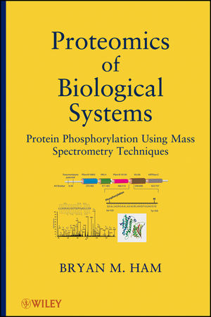
Proteomics of Biological Systems
John Wiley & Sons Inc (Verlag)
978-1-118-02896-4 (ISBN)
Phosphorylation is the addition of a phosphate (PO4) group to a protein or other organic molecule. Phosphorylation activates or deactivates many protein enzymes, causing or preventing the mechanisms of diseases such as cancer and diabetes. This book shows how to use mass spectrometry to determine whether or not a protein has been correctly modified by the addition of a phosphate group. It also provides a combination of detailed, step-by-step methodology for phosphoproteomic sample preparation, mass spectral instrumental analysis, and data interpretation approaches. Furthermore, it includes the use of bioinformatic Internet tools such as the Blast2GO gene ontology (GO) tool, used to help understand and interpret complex data collected in these studies.
BRYAN M. HAM, PhD, is a member of the American Society for Mass Spectrometry and the American Chemical Society. He has conducted proteomics and lipidomics research at The Ohio State University and Pacific Northwest National Laboratory in Richland, Washington. He is currently working for the Department of Homeland Security at the U.S. Customs and Border Protection New York Laboratory. He has published numerous research papers in peer-reviewed journals, and is the author of Even Electron Mass Spectrometry with Biomolecule Applications (Wiley).
Preface xvii Acknowledgments xxi
About the Author xxiii
1 Posttranslational Modification (PTM) of Proteins 1
1.1 Over 200 Forms of PTM of Proteins 1
1.2 Three Main Types of PTM Studied by MS 2
1.3 Overview of Nano-Electrospray/Nanofl ow LC-MS 2
1.3.1 Defi nition and Description of MS 2
1.3.2 Basic Design of Mass Analyzer Instrumentation 3
1.3.3 ESI 7
1.3.4 Nano-ESI 11
1.4 Overview of Nucleic Acids 15
1.5 Proteins and Proteomics 20
1.5.1 Introduction to Proteomics 20
1.5.2 Protein Structure and Chemistry 22
1.5.3 Bottom-Up Proteomics: MS of Peptides 27
1.5.3.1 History and Strategy 27
1.5.3.2 Protein Identifi cation through Product Ion Spectra 30
1.5.3.3 High-Energy Product Ions 36
1.5.3.4 De Novo Sequencing 37
1.5.3.5 Electron Capture Dissociation (ECD) 40
1.5.4 Top-Down Proteomics: MS of Intact Proteins 42
1.5.4.1 Background 42
1.5.4.2 GP Basicity and Protein Charging 42
1.5.4.3 Calculation of Charge State and Molecular Weight 44
1.5.4.4 Top-Down Protein Sequencing 46
1.5.5 Systems Biology and Bioinformatics 48
1.5.6 Biomarkers in Cancer 52
Reference 56
2 Glycosylation of Proteins 59
2.1 Production of a Glycoprotein 59
2.2 Biological Processes of Protein Glycosylation 59
2.3 N-Linked and O-Linked Glycosylation 60
2.4 Carbohydrates 60
2.4.1 Ionization of Oligosaccharides 64
2.4.2 Carbohydrate Fragmentation 65
2.4.3 Complex Oligosaccharide Structural Elucidation 70
2.5 Three Objectives in Studying Glycoproteins 72
2.6 Glycosylation Study Approaches 72
2.6.1 MS of Glycopeptides 73
2.6.2 Mass Pattern Recognition 75
2.6.2.1 High Galactose Glycosylation Pattern 75
2.6.3 Charge State Determination 76
2.6.4 Diagnostic Fragment Ions 76
2.6.5 High-Resolution/High-Mass Accuracy Measurement and Identification 76
2.6.6 Digested Bovine Fetuin 78
Reference 79
3 Sulfation of Proteins as Posttranslational Modification 81
3.1 Glycosaminoglycan Sulfation 81
3.2 Cellular Processes Involved in Sulfation 81
3.3 Brief Example of Phosphorylation 82
3.4 Sulfotransferase Class of Enzymes 82
3.5 Fragmentation Nomenclature for Carbohydrates 82
3.6 Sulfated Mucin Oligosaccharides 83
3.7 Tyrosine Sulfation 84
3.8 Tyrosylprotein Sulfotransferases TPST1 and TPST2 87
3.9 O-Sulfated Human Proteins 89
3.10 Sulfated Peptide Product Ion Spectra 89
3.11 Use of Higher Energy Collisions 93
3.12 Electron Capture Dissociation (ECD) 94
3.13 Sulfation versus Phosphorylation 95
Reference 97
4 Eukaryote PTM as Phosphorylation: Normal State Studies 99
4.1 Mass Spectral Measurement with Examples of HeLa Cell Phosphoproteome 99
4.1.1 Introduction 99
4.1.2 Protein Phosphatase and Kinase 99
4.1.3 Hydroxy-Amino Acid Phosphorylation 100
4.1.4 Traditional Phosphoproteomic Approaches 102
4.1.5 Current Approaches 103
4.1.5.1 Phosphoproteomic Enrichment Techniques 103
4.1.5.2 IMAC 104
4.1.5.3 MOAC 105
4.1.5.4 Methylation of Peptides prior to IMAC or MOAC Enrichment 107
4.1.6 The Ideal Approach 107
4.1.7 One-Dimensional (1-D) Sodium Dodecyl Sulfate (SDS) PAGE 108
4.1.8 Tandem MS Approach 108
4.1.8.1 pS Loss of Phosphate Group 109
4.1.8.2 pT Loss of Phosphate Group 112
4.1.8.3 pY Loss of Phosphate Group 113
4.1.9 Alternative Methods: Infrared Multiphoton Dissociation (IRMPD) and Electron Capture Dissociation (ECD) 115
4.1.10 Electron Transfer Dissociation (ETD) 115
4.2 The HeLa Cell Phosphoproteome 118
4.2.1 Introduction 118
4.2.2 Background of Study 118
4.2.3 What is Covered 119
4.2.4 Optimized Methods to Use for Phosphoproteomic Studies 119
4.2.4.1 Cell Culture 119
4.2.4.2 Extraction of HeLa Cell Proteins 120
4.2.4.3 Trizol Extraction and Tryptic Digestion 120
4.2.4.4 Solid-Phase Extraction (SPE) Desalting 120
4.2.4.5 Converting Peptide Carboxyl Moieties to Methyl Esters 121
4.2.4.6 Roche Complete Lysis-M, EDTA-Free Extraction 122
4.2.4.7 1-D SDS-PAGE Cleanup 122
4.2.4.8 In-Gel Reduction, Alkylation, Digestion, and Extraction of Peptides 122
4.2.4.9 Phosphopeptide Enrichment Using IMAC 123
4.2.5 Description of Instrumental Analyses 123
4.2.5.1 RP/Nano-HPLC Separation 123
4.2.5.2 MS Analysis 125
4.2.6 Current Approaches for Peptide Identification and False Discovery Rate (FDR) Determination 125
4.2.7 Results of the Protein Extraction and Preparation 126
4.2.7.1 Detergent Lysis, Trizol, and Ultracentrifugation 126
4.2.7.2 Nucleic Acid Removal with SDS-PAGE 127
4.2.8 HeLa Cell Phosphoproteome Methodology Comparison 128
4.2.8.1 Roche In-Solution versus Trizol Extraction 129
4.2.8.2 In-Solution and In-Gel Digests Phosphoproteome Coverage 129
4.2.9 Overall Conclusion 134
4.3 Nonphosphoproteome HeLa Cell Analysis 135
4.3.1 IMAC Flow Through Peptide Analysis 135
4.3.2 IMAC NaCl Wash Peptide Analysis 136
4.3.3 IMAC Flow Through versus NaCl Wash Comparison 138
4.3.4 Gene Ontology Comparison 138
4.3.5 IMAC Bed Nonspecifi c Binding Study 140
4.4 Reviewing Spectra Using the SpectrumLook Software Package 143
Reference 144
5 Eukaryote PTM as Phosphorylation: Perturbed State Studies 147
5.1 Study of the Phosphoproteome of HeLa Cells under Perturbed Conditions by Nano-High-Performance Liquid Chromatography HPLC Electrospray Ionization (ESI) Linear Ion Trap (LTQ)-FT/Mass Spectrometry (MS) 147
5.1.1 Introduction 147
5.1.2 Ataxia Telangiectasia Mutated (ATM) and ATM and Rad3-Related (ATR) 149
5.1.3 Background of Study 149
5.1.3.1 PP5 149
5.1.3.2 Functions of PP5 151
5.1.3.3 DDR of PP5 151
5.1.4 Review of Optimized Approach to Study 151
5.1.4.1 Producing Cell Cultures 151
5.1.4.2 Protein Extraction 152
5.1.4.3 Phosphopeptide Enrichment by IMAC 154
5.1.4.4 Reversed-Phase (RP)/Nano-HPLC Separation 155
5.1.4.5 LTQ-FT/MS/MS 156
5.1.4.6 Protein Identifi cation and False Discovery Rate (FDR) Determination 156
5.1.4.7 Phosphopeptide Quantitative Differential Comparison 157
5.1.4.8 Data Set Peak Matching and Alignment 157
5.1.4.9 Phosphopeptide Response Normalization 160
5.1.5 Phosphoproteome Gene Ontology (GO) Comparison 160
5.1.5.1 GO Cellular Component 162
5.1.6 Potential Regulated Target Proteins of PP5 162
5.1.6.1 Analysis of Variance (ANOVA) 162
5.1.6.2 Four Potential Target Proteins 166
5.1.7 GO Differential Comparison 167
5.1.7.1 GO Cellular Component 168
5.1.7.2 Infl uence of Classes or Categories of Proteins 168
5.1.7.3 Molecular Function Interacting Modules 168
5.1.8 Conclusion 175
5.1.9 Reviewing Spectra Using the SpectrumLook Software Package 175
Reference 176
6 Prokaryotic Phosphorylation of Serine, Threonine, and Tyrosine 181
6.1 Introduction 181
6.1.1 Serine (Ser)/Threonine (Thr)/Tyrosine (Tyr) Phosphorylation 181
6.1.2 Histidine (His) Phosphorylation 181
6.1.3 Caulobacter crescentus 181
6.1.4 Ser/Thr/Tyr Phosphorylation of C. crescentus 183
6.1.5 Ser/Thr/Tyr Phosphorylation of Bacillus subtilis and Escherichia coli 184
6.1.6 C. crescentus as Cell Cycle Model 185
6.1.7 Bacteria Starvation Response 187
6.1.8 First Coverage of C. crescentus Phosphoproteome 188
6.2 Optimized Methodology for Phospho Ser/Thr/Tyr Studies 188
6.2.1 Bacterial Strain and Growth Conditions 188
6.2.2 C. crescentus Cell Protein Extraction: Phosphoproteomics 189
6.2.3 Solid-Phase Extraction (SPE) Desalting 190
6.2.4 In Vitro Methylation of Peptides 190
6.2.5 Phosphopeptide Enrichment by IMAC 191
6.2.6 Normal Proteomics 192
6.2.7 pY Enrichment by IP 192
6.2.8 RP/Nano-High-Performance Liquid Chromatography (HPLC) Separation 192
6.2.9 LC-Linear Ion Trap (LTQ)-Orbitrap MS/MS 193
6.2.10 LTQ-Fourier Transform (FT)/MS/MS 193
6.2.11 Peptide Identification and False Discovery Rate (FDR) Determination 193
6.2.12 Peptide Quantitative Comparison 194
6.3 Identifi cation of the Components of the Ser/Thr/Tyr Phosphoproteome in C. crescentus Grown in the Presence and Absence of Glucose 194
6.3.1 Total Phosphoprotein Identifications 194
6.3.2 MSA Spectra 196
6.3.3 Phosphorylation Sites Identifi ed 196
6.3.4 Ser/Thr/Tyr Phosphoproteome of C. crescentus 205
6.3.5 Phosphorylated His and Aspartate 213
6.3.6 Cell Cycle His Kinase CckA 215
6.3.7 Phosphoglutamate 216
6.3.8 Enriched Tyr Phosphoproteome of C. crescentus 216
6.3.8.1 Sensor His Kinase KdpD 216
6.3.8.2 TonB-Dependent Receptor Proteins 216
6.3.9 Carbon Environment-Shared Phosphoproteome 217
6.3.9.1 Two-Component His Kinases 217
6.3.9.2 Multiply Phosphorylated Kinases 217
6.3.9.3 pTPLAALpSAQSRRAR Peptide as Sensor His Kinase 217
6.3.9.4 Aspartate Phosphorylated Tyr Kinase DivL 217
6.3.10 Carbon-Rich versus Carbon-Starved Class/Category 225
6.3.10.1 Localization of Phosphoproteome of C. crescentus 225
6.3.10.2 Integral Membrane Proteins 225
6.3.10.3 Function of Phosphoproteome of C. crescentus 225
6.3.11 Carbon-Rich versus Carbon-Starved Unique Phosphorylated Proteins 227
6.3.11.1 Carbon-Rich Environment Phosphorylated Proteins 227
6.3.11.2 Carbon-Starved Environment Phosphorylated Proteins 227
6.3.11.3 Decreased Normal Activity 232
6.3.12 Confi rmation of Decreased Energy Pathways 232
6.3.12.1 Carbon-Rich Mitochondrial Localization 232
6.3.12.2 Normal Proteome Glycolytic Pathway 233
6.3.12.3 Starvation Survival Response 233
6.3.13 Phosphopeptide Quantitative Differential Comparison 233
6.3.13.1 Upregulation in Phosphorylation 234
6.3.13.2 Adaptive Response with Phosphorylation 234
6.3.13.3 Upregulation NAD-Dependent GDH 234
6.3.13.4 Downregulation of Flagellin Protein 235
6.3.14 Carbon-Rich versus Carbon-Starved Normal Proteome Time Course Study 235
6.3.14.1 Entire Proteome Localization and Function 235
6.3.14.2 Regulated Proteins 237
6.3.14.3 Localization of Regulated Proteins 237
6.3.14.4 Function of Regulated Proteins 238
6.3.14.5 Normal Proteome Energy Pathways 239
6.3.14.6 Overlap of Phosphorylated Proteins and Regulated Normal Proteome 239
6.3.14.7 Differences of Phosphorylated Proteins 240
6.3.14.8 Localization of Phosphorylated Proteins 240
6.3.14.9 Direct Relationships Observed 240
6.3.15 Conclusions 243
6.3.16 Supplementary Material 243
6.3.16.1 Reviewing Spectra Using the SpectrumLook Software Package 243
Reference 244
7 Prokaryotic Phosphorylation of Histidine 249
7.1 Phosphohistidine as Posttranslational Modification (PTM) 249
7.2 Bacterial Kinases and the Two-Component System 250
7.3 Measurement of Phosphorylated His (pH) 251
7.3.1 Stabilities of Phosphorylated Amino Acids 251
7.3.2 Immobilized Metal Affinity Chromatography (IMAC) and Mass Spectrometry (MS) 252
7.4 In Vitro and In Vivo Study of pH-Containing Peptides by Nano-ESI Tandem MS 255
7.4.1 Introduction 255
7.4.2 Background of Study 257
7.4.2.1 Bacteria Models of Ser/Thr/Tyr Phosphorylation 257
7.4.2.2 Prokaryotic Phosphorylation of His 258
7.4.2.3 C. crescentus 258
7.4.2.4 Mass Spectral Measurement of Phosphohistidine 258
7.4.3 Optimized Methodology for Phosphohistidine Studies 259
7.4.3.1 In Vitro Selective pHis Phosphorylation 259
7.4.3.2 In Vitro Phosphorylation of Angio II (Sar1Thr8) 261
7.4.3.3 In Vitro Methylation of Peptides 262
7.4.3.4 C. crescentus Cell Protein Extraction with V-8 Protease Digestion 262
7.4.3.5 1-D SDS-Polyacrylamide Gel Electrophoresis (PAGE) 263
7.4.3.6 Phosphohistidine Enrichment by Cu(II)-Based IMAC 264
7.4.3.7 Reversed-Phase (RP)/Nano-HPLC Separation 265
7.4.3.8 Nano-ESI Nano-HPLC MS 266
7.4.3.9 Peptide Identification and False Discovery Rate (FDR) Determination 268
7.4.4 C18 RP LC Behavior 268
7.4.5 Phosphohistidine Loses HPO3 and H3PO4 270
7.4.5.1 Rational for H3PO4 Loss 272
7.4.6 Q-TOF/MS/MS Product Ion Spectra 277
7.4.6.1 pH-Containing Peptide INpHDLR 277
7.4.6.2 Doubly Charged (2+) Peptide INpHDLR 279
7.4.6.3 pH-Containing Peptide pHLGLAR 279
7.4.6.4 Singly Charged (1+) Peptide pHLGLAR 280
7.4.7 Behavior of Monophosphohistidine and Diphosphohistidine Peptide 281
7.4.7.1 Peptide Angio I as DRVYIHPFHL 281
7.4.8 Behavior of Phosphotyrosine and Phosphohistidine Peptide 285
7.4.8.1 Peptide Angio II as DRVpYIHPF 285
7.4.8.2 Phosphorylated Angio II as DRVpYIpHPF 285
7.4.9 Behavior of Phosphotyrosine-, Phosphothreonine-, and Phosphohistidine-Containing Peptide 287
7.4.9.1 Peptide Angio II (Sar1Thr8) 287
7.4.10 Validation of Cu(II)-Based IMAC Phosphohistidine Enrichment 291
7.4.10.1 Fe(III)-Based IMAC versus Cu(II) Based 292
7.4.10.2 Cu(II)-Based IMAC of Angio I 292
7.4.10.3 Cu(II)-Based IMAC of Angio II 293
7.4.11 In Vivo Measurement of Phosphohistidine 293
7.4.11.1 Time-Based Digestion Study 293
7.4.11.2 Phosphohistidine-Containing Peptides 294
7.4.11.3 Phosphohistidine Product Ion Spectra 294
7.4.12 Gene Ontology of Phosphorylated Proteins 296
7.4.12.1 Localization of Phosphorylated Proteins 296
7.4.12.2 Function of Phosphorylated Proteins 304
7.4.13 Predicted Regulatory Protein Motif Study 307
7.4.14 Validation of Phosphohistidine-Containing Proteins 308
7.4.14.1 Phosphorylation Motif Study 308
7.4.14.2 Phosphohistidine Kinase Motif 309
7.4.15 The pDpH Motif 310
7.4.16 Conclusions 311
7.5 Supplementary Material 311
7.5.1 Reviewing Spectra Using the SpectrumLook Software Package 311
Reference 313
Appendix I Atomic Weights and Isotopic Compositions 317
Appendix II Periodic Table of the Elements 325
Appendix III Fundamental Physical Constants 327
Glossary 329
Index 345
| Erscheint lt. Verlag | 16.12.2011 |
|---|---|
| Verlagsort | New York |
| Sprache | englisch |
| Maße | 160 x 236 mm |
| Gewicht | 658 g |
| Themenwelt | Naturwissenschaften ► Biologie ► Biochemie |
| Naturwissenschaften ► Chemie ► Analytische Chemie | |
| ISBN-10 | 1-118-02896-1 / 1118028961 |
| ISBN-13 | 978-1-118-02896-4 / 9781118028964 |
| Zustand | Neuware |
| Haben Sie eine Frage zum Produkt? |
aus dem Bereich


