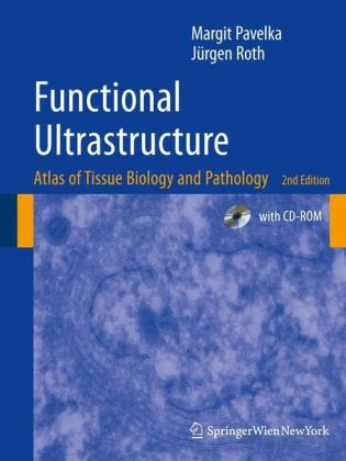
Functional Ultrastructure
Springer Wien (Verlag)
978-3-211-99389-7 (ISBN)
- Titel erscheint in neuer Auflage
- Artikel merken
The period between 1950 and 1980 were the golden unique insights into how pathological processes affect years of transmission electron microscopy and produced cell organization. a plethora of new information on the structure of cells This information is vital to current work in which that was coupled to and followed by biochemical and the emphasis is on integrating approaches from functional studies. TEM was king and each micrograph proteomics, molecular biology, genetics, genomics, of a new object produced new information that led to molecular imaging and physiology and pathology to novel insights on cell and tissue organization and their understand cell functions and derangements in disease. functions. The quality of data represented by the images In this current era, there is a growing tendency to of cell and tissues had been perfected to a very high level substitut e modern light microscopic techniques for by the great microscopists of that era including Palade, electron microscopy, because it is less technically Porter, Fawcett, Sjostrand, Rhodin and many others. At demanding and is more readily available to researchers- present, the images that we see in leading journals for This atlas reminds us that the information obtained by the most part do not reach the same technical level and electron microscopy is invaluable and has no substitute.
Professor Margit Pavelka, MD. Studies in Medicine, University of Vienna. Medical training at the Vienna Hospital “Rudolfstiftung” and at the Vienna General Hospital. Specialization in Internal Medicine. Resident at the Institute of Micromorphology and Electron Microscopy, Vienna. Specialization in the fields of electron microscopy, cytology, histochemistry, cytochemistry, and ultrastructural histology.Habilitation and University Docent in Histology and Embryology. Professor of Histology and Embryology, University of Innsbruck. Currently Professor of Histology and Embryology, Medical University of Vienna. Main research interests: Morpho-functional organization of the Golgi apparatus; secretory and endocytic pathways; visualization of cellular dynamics; cell traffic in health and disease; cellular stress. Professor Jürgen Roth, MD, PhD. Studies in Medicine, Friedrich-Schiller-University, Jena. Resident at the Institute of Pathology, Friedrich-Schiller-University, Jena. Habilitation and University Docent in Pathology, Friedrich-Schiller-University, Jena. Research Associate, Department of Morphology, University of Geneva. Associate Professor of Cell Biology, Biozentrum, University of Basel. Professor Emeritus of Cell and Molecular Pathology, University of Zurich. Currently Distinguished Professor, World Class University Programme, Department of Biomedical Science, Yonsei University, Seoul. Main research interests: Topography of protein glycosylation; protein quality control; protein folding diseases; carcinoma-associated glycoconjugates.
The Cell.- Structural Organisation of a Mammalian Cell.- Architecture of the Cell Nucleus.- Cytochemical Detection of Ribonucleoproteins.- Nuclear Lamina.- Detection of Sites of NA Replication and of Interphase Chromosome domains.- Nucleolus.- Changes of the Nucleolar Architecture.- Detection of Sites of RNA Synthesis.- Nuclear Pore Complexes.- Nuclear Pore Complexes: Structural Changes as Monitored by Time-Lapse Atomic Force Microscopy.- Mitosis and Cell Division.- Apoptosis.- Viral Inclusions.- Secretory Pathway of Pancreatic Acinar Cells.- Endomembrane System of Dinoflagellates.- Ribosomes, Rough Endoplasmic Reticulum.- Nuclear Envelope and Rough Endoplasmic Reticulum.- Annulate Lamellae.- Rough Endoplasmic Reticulum: Site of Protein Translocation and Initiation of Protein N-Glycosylation.- Oligosaccharide Trimming, Reglucosylation, and Protein Quality Control in the Rough Endoplasmic Reticulum.- Rough Endoplasmic Reticulum: Storage Site of Aggregates of Misfolded Glycoproteins.- Russell Bodies and Aggresomes Represent Different Types of Protein Inclusion Bodies.- Smooth Endoplasmic Reticulum.- Proliferation of the Smooth Endoplasmic Reticulum.- Pre-Golgi Intermediates.- Pre-Golgi Intermediates: Oligosaccharide Trimming and Protein Quality control.- Golgi Apparatus: A Main Crossroads Along Secretory Pathways.- Protein Secretion Visualised by Immunoelectron Microscopy.- Protein N-Glycosylation: Oligosaccharide Trimming in the Golgi Apparatus and Pre-Golgi Intermediates.- Golgo Apparatus: Site of Maturation of Aspara Gine-Linked Oligo Saccharides.- Cell Type-Related Variations in the Topography of Golgi Apparatus Glycosylation Reactions.- Cell Type-Related Differences in Oligosaccharide Structure.- Topography of Biosynthesis of Serine/Threonine-Linked Oligosaccharides.- Golgi Apparatus and TGN — Structural Considerations.- Golgi Apparatus and TGN — Secretion and Endocytosis.- Golgi Apparatus, TGN and Trans Golgi-ER.- Golgi Apparatus, TGN and Trans Golgi-ER: Tilt Series.- Structure of the TGN.- Brefeldin A-Induced Golgi Apparatus Disassembly.- Brefeldin A-Treatment: Tubulation of Golgi Apparatus and Endosomes.- Brefeldin A-Treatment: Effect on Retrograde Transport of Internalised WGA.- Brefeldin A-Treatment: Transitional ER-Elements and Pre-Golgi Intermediates.- Heat Shock Response of the Golgi Apparatus.- Golgi Apparatus Changes Upon ATP-Depletion and ATP-Replenishment.- Secretory Granules.- Secretory Granule Types.- Goblet Cell — Compound Exocytosis.- Receptor-Mediated Endocytosis Via Clathrin-Coated Vesicles and Virus Endocytosis.- Endosomes and Endocytic Pathways.- Endocytic Trans Golgi Network and Retrograde Traffic into the Golgi Apparatus.- Tubular Pericentriolar Endosomes.- Langerhans Cells and Birbeck Granules: Antigen Presenting Dendritic Cells of the Epidermis.- Caveolae.- Fluid-Phase Endocytosis and Phagocytosis.- Lysosomes.- Lysosomes: Localisation of Acid Phosphatase, Lamp and Polylactosamine.- I-Cell Disease.- Gaucher’s disease.- Fabry’s Disease.- GM2 Gangliosidoses.- Farber’s Disease.- Wolman’s Disease.- Glycogenosis Type II.- Cystinosis.- Autophagy: Limited Self-Digestion.- Pexophagy: Autophagy of Peroxisomes.- Mitochondria: Crista and Tubulus Types.- Abnormalities of Mitochondria.- Peroxisomes: Multitalented Organelles.- Peroxisome Biogenesis.- Peroxisomes: Adaptive Changes.- Peroxisomal Disorders.- Glycogen.- Glycogenosis Type I.- Erythropoietic Protoporphyria.- Cytocentre, Centrosome, and Microtubules.- Effects of Microtubule Disruption.- Actin Filaments.- Intermediate Filaments.- Mallory Bodies.- The Plasma Membrane.- Cells in Culture.- Brush Cell.- Glycocalyx (Cell Coat).- Glycocalyx: Cell Type Specificity and Domains.- Glycocalyx Changes in Tumours.- Junctional Complex.- Tight Junctions and Gap Junctions.- Tunneling Nanotubes.- Spot Desmosomes.- Selectin — Ligand-Mediated Cell-Cell Interaction.- Cellular Interdigitations.- Basal Labyrinth.- Basement Membrane.- Glomerular Basement Membrane.- Alport’S Syndrome (Hereditary Nephritis).- Descemet’s Membrane.- Skin Basement Membrane and Keratinocyte Hemidesmosomes: An Epithel-Connective Tissue Junctional Complex.- Epidermolysis Bullosa Simplex.- Principles of Tissue Organisation.- Pancreatic Acinus.- Acinar Centre: Acinar and Centroacinar Cells.- Pancreatic Intercalated Duct.- Submandibular Gland.- Goblet Cells — Unicellular Glands.- Parietal Cells of Stomach: Secretion of Acid.- Intercalated Cells of Kidney: Important Regulators of Acid-Base Balance.- Endocrine Secretion: Insulin-Producing Beta Cells of Islets of Langerhans.- Impaired Insulin Processing in Human Insulinoma.- Cells of the Disseminated Endocrine System.- Liver.- Liver: Hepatocytes, Kupffer cell, Cell of Ito.- Liver Epithelium: Bile Canaliculi.- Liver Epithelium: Pathway of Secretory Lipoprotein Particles.- Congenital Hepatic Fibrosis.- Choroid Plexus Ependyma.- Small Intestine: Absorptive Cells.- Small Intestine: Pathway of Lipids.- Renal Proximal Tubule: A Reabsorption Plant.- Parathyroid Hormone Response of Renal Proximal Tubules.- Photoreceptor Cells of the Retina: Signalling of Light.- Photoreceptor Cells of the Retina: Light-Induced Apoptosis.- Olfactory Epithelium.- Corneal Epithelium.- Epidermis.- Differentiation of Keratinocytes and Formation of the Epidermal Fluid Barrier.- The Tracheo-Bronchial Epithelium.- Ciliary Pathology: Immotile Cilia Syndrome and Kartagener Syndrome.- Alveoli: Gas Exchange and Host Defense.- Umbrella Cell — Surface Specialisations.- Umbrella Cell — Fusiform Vesicles.- Continuous Capillary, Weibel-Palade Bodies.- Hyaline Arteriolosclerosis.- Fenestrated Capillary.- Endothelio-Pericyte and Endothelio-Smooth Muscle Cell Interactions.- Glomerulus: A Specialised Device for Filtering.- Pathology of the Glomerular Filter: Minimal Change Glomerulopathy and Congenital Nephrotic Syndromes.- Pathology of the Glomerulus: Membranous Glomerulonephritis.- Pathology of the Glomerulus: Membranoproliferative Glomerulonephritis.- Pathology of the Glomerulus: IgA Nephropathy (Berger).- Pathology of the Glomerulus: Chronic Allograft Glomerulopathy.- Loose Connective Tissue.- Fibroblast, Fibrocyte, Macrophage.- Collagen and Elastic Fibres.- Eosinophilic Granulocyte, Plasma Cell, Macrophage, Mast Cell.- Dense Connective Tissue: Collagen Bundles in the Cornea.- Bowman’s Layer.- Amyloidosis of Kidney.- Amyloid Fibrils: Growth as Seen by Time Lapse, Atomic Force Microscopy.- White Adipose Tissue.- Brown Adipose Tissue.- Articular Cartilage.- Osteoblasts and Osteocytes.- Osteoclast.- Myofibrils and Sarcomere.- Sarcoplasmic Reticulum, Triad, Satellite Cell.- Neuromuscular Junction.- Muscular Dystrophies.- Glycogenosis Type II (Pompe).- Myofibrils, Intercalated Disk.- Smooth Muscle Cells, Synapse Á Distance.- CADASIL.- Central Nervous System: Neuron, Glial Cells.- Blood-Brain Barrier, Synapses.- Structure of the Synaptic Terminal.- Unmyelinated Nerve Fibre.- Peripheral Nerve: Connective Tissue Components.- Myelinated Nerve Fibre, Myelin.- Node of Ranvier.- Axonal Degeneration.- Neuroaxonal Dystrophy.- Neuropathies Associated with Dysproteinaemias.- Metachromatic Leukodystrophy.- Neuronal Ceroid Lipofuscinosis.- Red Blood Cells and Cells of the Erythroid Lineage.- Neutrophilic Granulocyte.- Eosinophilic Granulocyte.- Basophilic Granulocyte.- Monocyte.- Lymphocyte.- Megakaryocyte and Thrombocyte.- Thrombocytes.
| Sprache | englisch |
|---|---|
| Maße | 210 x 277 mm |
| Gewicht | 1415 g |
| Einbandart | gebunden |
| Themenwelt | Naturwissenschaften ► Biologie ► Mikrobiologie / Immunologie |
| Schlagworte | biosynthesis • Cell • cell architecture • Cell Biology • cell nucleus • Cell organelles • Cells • cell structure • cytology • electron microscopy • Endoplasmatisches Reticulum • Glycogen • golgi apparatus • Histology • Matrix • Molecular Biology • Pathology • proteins • RNA • tissue • ultrastructural pathology • Ultrastructure • Zelle (biolog.) • Zelle (Biologie) |
| ISBN-10 | 3-211-99389-4 / 3211993894 |
| ISBN-13 | 978-3-211-99389-7 / 9783211993897 |
| Zustand | Neuware |
| Haben Sie eine Frage zum Produkt? |
aus dem Bereich


