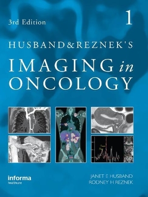
Husband and Reznek's Imaging in Oncology, Third Edition
CRC Press (Verlag)
978-0-415-45167-3 (ISBN)
- Titel erscheint in neuer Auflage
- Artikel merken
In recent years there has been recognition of the central role of imaging in the management of patients with cancer. The third edition of this widely acclaimed book builds on the foundations laid down by the first edition, the 1998 winner of the Royal Society's award for the Multi-author Textbook of the Year, and the second (2004). The core of the book deals with the application of imaging in all tumours. An extensively referenced, evidence-based analysis is given of the role of imaging in planning treatment. Experienced opinion is provided as to the advantages and limitations of all relevant imaging modalities including ultrasound, CT, MRI, PET/CT and other nuclear medicine techniques.
Imaging in the recognition of metastatic disease is covered in detail with chapters detailing the range of appearances of deposits in each organ. Much attention is given to the differential diagnosis of focal pathology in each organ in patients with underlying malignancy and useful protocols for the performance of the study provided.
While including a section on new horizons in cancer imaging, such as the imaging of angiogenesis, MR lymphography and the rapidly evolving field of molecular imaging, the editors have not neglected the more traditional general principles of cancer behaviour and imaging. Hence, an outline of cancer surgery, chemotherapy and radiotherapy is provided by experts. As therapy becomes more successful, increasingly important issues for the radiologist such as the assessment of response, the effect of treatment on normal tissues, the complications of treatment, and the risk of second malignancies are presented.
There are several outstanding features to this book: the colour diagrams of the staging systems are exquisite and allow the often complex systems to be understood easily and memorized; the organization of the book into different, clearly defined sections allow it to be used not only as a reliable reference text, but also to be read easily; key points and summaries in point form are provided throughout for quick revision.
This comprehensive book will not only be essential reading for all radiologists, but also important for all members of a multidisciplinary team looking after patients with cancer.
Preface Foreword from the 2nd edition Foreword for the 3rd edition PART I - GENERAL PRINCIPLES 1. Trends in cancer incidence, survival and mortality 2. Staging of Cancer 3. Multidisciplinary treatment of cancer: Surgical 4. Multidisciplinary treatment of cancer: Chemptherapy 5. Multidisciplinary treatment of cancer: Radiotherapy 6. Assesment of Response to Treatment 7. Second maliginacies PART II - PRIMARY TUMOUR EVALUATION AND STAGING 8. Lung cancer 9. Mediastinal Tumours 10. Pleaural tumours 11. Oesphogeal Cancer 12. Gastric Cancer 13. Colorectal Cancer 14. Primary Tumours of the Liver and Biliary Tract 15. Renal Tumours 16. Primary Adrenal Malignancy 17. Pancreatic Malignancy 18. Bladder Cancer 19. Prostate Cancer 20. Testicular germ cell tumours 21. Ovarian Cancer 22. Uterine and Cervical Tumours 23. Primary Retroperitoneal Tumours 24. Primary Bone Tumours 25. Soft Tissue Sarcomas 26. Breast Cancer 27. Paranasal Sinus Neoplasms 28. Tumours of the Pharynx, Tongue and Mouth 29. Laryngeal Tumours 30. Thyroid Cancer 31. Primary Tumours of the Central Nervous System 32. Neuroendocrine Tumours PART III - HAEMATOLOGY MALIGNANCY 33. Lymphoma 34. Multiple Myeloma 35. Leukaemia PART IV – PAEDIATRICS 36. General Principles in Paediatric Oncology 37. Wilm's Tumour and Associated Neoplasms of the Kidney 38. Neuroblastoma 39. Uncommon Paediatric Neoplasms PART V – METASTASES 40. Lymph node Metastases 41. Lung and Pleural Metastases 42. Bone Metastases 43. Liver Metastases 44. Metastatic effects on the Nervous System 45. Adrenal Metastases 46. Peritoneal Metastases 47. Spleen 48. Malignant Tumours of the Skin 49. Radiological Investigation of Carcinoma of Unknown Primary Site PART VI - IMAGING AND TREATMENT 50. Interventional Imaging: General Applilcations 51. Interventional Imaging: Tumour Ablation 52. Imaging for Radiotherapy Treatment Planning 53. Radiological Manifestations of Acute Complications of Treatment PART VII - EFFECTS OF TREATMENT ON NORMAL TISSUE 54. Effects of Treatment on Normal Tissue: Thorax 55. Bone and Bone Marrow 56. Abdomen and Pelvis PART VIII - THE IMMUNOCOMPROMISED HOST 57. The Immunocompromised Host: Clinical Considerations 58. The Immunocompromised: Central Nervous System 59. The Immunocompromised Host: Chest 60. The Immunocompromised Host: Abdomen and Pelvis PART IX - FUNCTIONAL IMAGING 61. Positron Emission Tomography - Principles and Clinical Applications 62. Functional Imaging: Clincal Applications in Molecular Targeted Therapy 63. Measurement of Angiogensis- MRI Principles & Practice 64. Measurement of angiogenesis - CT principles and practice 65. Magnetic resonance: Emerging technologies and applications
| Erscheint lt. Verlag | 23.12.2009 |
|---|---|
| Zusatzinfo | 1100 Halftones, color; 1400 Halftones, black and white |
| Verlagsort | London |
| Sprache | englisch |
| Maße | 214 x 285 mm |
| Gewicht | 5511 g |
| Themenwelt | Medizin / Pharmazie ► Medizinische Fachgebiete ► Onkologie |
| Medizinische Fachgebiete ► Radiologie / Bildgebende Verfahren ► Radiologie | |
| ISBN-10 | 0-415-45167-1 / 0415451671 |
| ISBN-13 | 978-0-415-45167-3 / 9780415451673 |
| Zustand | Neuware |
| Informationen gemäß Produktsicherheitsverordnung (GPSR) | |
| Haben Sie eine Frage zum Produkt? |
aus dem Bereich


