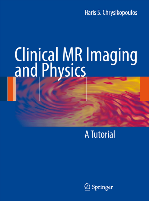
Clinical MR Imaging and Physics
Springer Berlin (Verlag)
978-3-540-77999-5 (ISBN)
Resonance.- Electromagnetic Fields.- Macroscopic Magnetization.- Macroscopic Magnetization Revisited.- Excitation Phenomena.- T1 Relaxation (Longitudinal or Spin-Lattice Relaxation).- T2 Relaxation (Transverse or Spin Spin Relaxation).- Magnetic Substrates of T1 Relaxation.- Magnetic Substrates of T2 Relaxation.- Proton (Spin) Density Contrast.- Partial Saturation.- Free Induction Decay.- Spin Echo.- Integration of T1, T2, and Proton Density Phenomena.- Inversion Recovery.- Image Formation Fourier Transform Gradients.- Gradient Echo Imaging.- Pulse Sequences.- Fast or Turbo Spin Echo Imaging.- Selective Fat Suppression.- Chemical Shift Imaging.- Magnetization Transfer Contrast.- Diffusion.- Artifacts.- Noise.- Imaging Time.- Resolution.- Contrast Agents.- Blood Flow.- MR Angiography.- Basics of MR Examinations and Interpretation.
From the reviews:
"This introductory book is designed to take readers from the fundamentals of proton interactions to the interpretation of clinical MR imaging studies. ... All healthcare professionals are the intended audience. The book is certainly suitable for radiologists interested in MRI or MR technologists wanting to learn the essentials of the technique. ... provides a good starting point for those involved in MR imaging with a clinical perspective. It is well-illustrated and practically demonstrates how physical principles directly govern the images of any MR imaging study." (R. Terry Thompson, Doody's Review Service, March, 2009)
| Erscheint lt. Verlag | 20.11.2008 |
|---|---|
| Zusatzinfo | IX, 176 p. |
| Verlagsort | Berlin |
| Sprache | englisch |
| Maße | 203 x 276 mm |
| Gewicht | 596 g |
| Themenwelt | Medizinische Fachgebiete ► Radiologie / Bildgebende Verfahren ► Kernspintomographie (MRT) |
| Medizinische Fachgebiete ► Radiologie / Bildgebende Verfahren ► Radiologie | |
| Schlagworte | Angiography • Diagnosis • Hardcover, Softcover / Medizin/Klinische Fächer • HC/Medizin/Klinische Fächer • Kernresonanztomographie • Magnetic Resonance • Magnetic Resonance Imaging • Magnetic Resonance Imaging (MRI) • MR Angiography • MR imaging • MR Physics |
| ISBN-10 | 3-540-77999-X / 354077999X |
| ISBN-13 | 978-3-540-77999-5 / 9783540779995 |
| Zustand | Neuware |
| Informationen gemäß Produktsicherheitsverordnung (GPSR) | |
| Haben Sie eine Frage zum Produkt? |
aus dem Bereich


