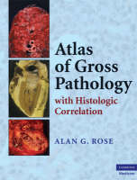
Atlas of Gross Pathology
With Histologic Correlation
Seiten
2008
Cambridge University Press (Verlag)
978-0-521-86879-2 (ISBN)
Cambridge University Press (Verlag)
978-0-521-86879-2 (ISBN)
- Titel ist leider vergriffen;
keine Neuauflage - Artikel merken
This atlas provides a comprehensive illustration and description of a wide range and number of pathologic processes and diseases affecting all major organs of the body. Histologic illustrations of selected gross lesions are also included where relevant. The book is illustrated with more than 1,200 color photomicrographs.
Detailed understanding of gross pathology is mandatory for successful pathologists, but this knowledge also provides a sound foundation for those intending to become surgeons, internists, and obstetrician/gynecologists. For pathologists, pathology residents, and pathology assistants, knowledge of gross pathology is essential for guidance in selecting the correct areas of pathologic lesions to sample for microscopy and frozen section examination. This atlas provides a comprehensive illustration and description of a wide range and number of pathologic processes and diseases affecting all the major organs of the body. Emphasis is placed on how the anatomic structure of different organs may determine the pattern of involvement by disease processes and how such patterns may aid in the correct diagnosis of the gross pathology. In some cases, multiple illustrations of disease processes are given to show evolution of the disease. Histologic illustrations of selected gross lesions are also included where relevant. The atlas is illustrated with more than 1,200 color photomicrographs.
Detailed understanding of gross pathology is mandatory for successful pathologists, but this knowledge also provides a sound foundation for those intending to become surgeons, internists, and obstetrician/gynecologists. For pathologists, pathology residents, and pathology assistants, knowledge of gross pathology is essential for guidance in selecting the correct areas of pathologic lesions to sample for microscopy and frozen section examination. This atlas provides a comprehensive illustration and description of a wide range and number of pathologic processes and diseases affecting all the major organs of the body. Emphasis is placed on how the anatomic structure of different organs may determine the pattern of involvement by disease processes and how such patterns may aid in the correct diagnosis of the gross pathology. In some cases, multiple illustrations of disease processes are given to show evolution of the disease. Histologic illustrations of selected gross lesions are also included where relevant. The atlas is illustrated with more than 1,200 color photomicrographs.
Dr Alan G. Rose, MD, FRCPath, FACC, is Professor of Pathology at the University of Minnesota Medical School. He is also the Director of Autopsy Service at the University of Minnesota Medical Center, Fairview; the Residency and Fellowship Training Program; and the Medical School Pathology Teaching Program at the University of Minnesota.
1. Cardiac diseases; 2. Pulmonary pathology; 3. Kidneys, ureters, and urinary bladder; 4. Liver, biliary system, and pancreas; 5. Salivary glands and GIT; 6. Female genital tract and breast; 7. Diseases of the male genital system; 8. Bones and joints; 9. Spleen, lymph nodes, and thymus; 10. Endocrine system; 11. Nervous system.
| Zusatzinfo | 130 Plates, color |
|---|---|
| Verlagsort | Cambridge |
| Sprache | englisch |
| Maße | 226 x 286 mm |
| Gewicht | 2900 g |
| Themenwelt | Studium ► 2. Studienabschnitt (Klinik) ► Pathologie |
| ISBN-10 | 0-521-86879-3 / 0521868793 |
| ISBN-13 | 978-0-521-86879-2 / 9780521868792 |
| Zustand | Neuware |
| Informationen gemäß Produktsicherheitsverordnung (GPSR) | |
| Haben Sie eine Frage zum Produkt? |
Mehr entdecken
aus dem Bereich
aus dem Bereich


